
Are you struggling with low back pain despite having tried “everything”? Many feel like this, but reality is that they haven’t tried everything — and many have done the wrong things despite their efforts being based on well-meaning advice. This, of course, is not the patient’s fault. Unfortunately, medical research is torn pertaining to the causes of chronic pain, and generally suggests in its consensus that it is psychological (Engelmann 2026, Lederman 2011). World Health Organization states that a staggering 85% of all LBP is considered idiopathic.(WHO) Many treatment modalities are therefore based on ameliorating the symptoms, as they believe they cannot find the cause (although they will rarely admit this to the patient). This article will discuss the most common causes for stubborn low back pain.
Generally, low back pain tends to originate from one or more out of the following six:
- Spinogenic pain, ie. the pain is originating from the spine and its related anatomy; the vertebrae and discs
- Sacroiliac joint pain
- Radiculopathic pain, ie. pain originating from a pinched nerve root in the spine
- Myogenic pain, ie. the pain is originating from muscles in the low back area
- Neurogenic pain, ie. the pain is originating from peripheral nerves being entrapped outside of the spine, usually by muscle tissue
- Clenching issues
Many nuances and caveats will come into play when determining not only which of these that may be affecting you, but also how to treat it.
Spinogenic and radiculopathic pain
The first step is to identify whether or not the patient’s symptoms are suggestive of a spinogenic condition. For patients with spine-related pain, we would typically expect those symptoms to be elicited through provocation of the low spine, and you would expect the symptoms to be situated along the spine itself, or within the expected pain zones for a pinched nerve root. This, naturally, requires some understanding of anatomy, but I will try to provide the basics.
Firstly, can you invoke the symptoms by repeated bending forward, backward or to the sides? If you can, is the pain located along the spine itself, or is it located to the side of the spine? Does the pain radiate into the buttock or down the leg? If your symptoms are 1. elicited with spinal provocation (repeated movement or loaded movement, eg. bending over and lifting something heavy) 2. elicits pain in the spine itself and not outside of the spine, or 3. elicits pain along one of the nerve root-related dermatomes, then you are probably dealing with a spine problem! If there is a presence of mild radiculopathy, then a Lasegue’s or Reversed Lasegue’s Test may be positive as well.
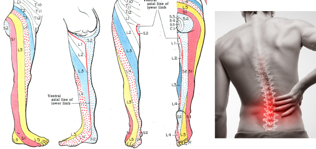
Fig. 1: Left) Dermatomes of the lower extremity. Beware that radiculopathy does NOT have to extend into the entire dermatome; it can sometimes be felt just in the buttock, in the calf or behind the knee, etc. It varies from case to case. Right) Spine-related pain (coming from the disc or vertebra) would be expected to predominantly manifest in, or along the spinal column itself.
Persistent or recurrent cases of spinogenic pain can have various causes. In cases of longstanding trouble, I always ask for an MRI of the lumbosacral spine (low back). Today’s consensus is that an MRI is not needed unless you are suspecting a dangerous lesion, eg. something that can cause permanent damage to your nerves. I disagree with this; an MRI is also incredibly useful in determining prognosis (expected outcome) — information of essential value to both the patient and caregiver alike. The main task here, is to ensure that there are no lesions present in the imaging (in your spine) that would prevent good outcomes with conservative management, and I will get back to what these findings are in a general sense a little later.
Presuming an MRI is either negative or shows only normal wear without any red or yellow flags, and within the scope of spinogenic pain, then you are most likely dealing with repeated irritation of the discs. This is a controversial topic, although it really shouldn’t be. Lots and lots of research has shown that flexion (bending) of the spine will ultimately lead to a herniated disc (Potvin 1991; Marshall 2010 Aultman 2004; Veres 2009; McGill 2000; Tampier 2007; Wade 2014). Some have said, in protest, that if we can’t bend the spine then there’s nothing we can do, and studied the psycho-social aspects of pain instead, finding that most people with chronic pain have proclivities to depression, anxiety, etc (Landmark 2024). The scope of this article is not to discuss all of these controversies, but rather to provide practical guidestones for the reader. I have gone in-depth in these controversies in previously published research (Larsen, 2018).
Repeated flexion of the spine will damage the spine. However, what most people do not consider here is the notion, fact, that spinal bending can be executed posturally, meaning that the patient can be standing and sitting in a way that constantly flexes and wears the spine without knowing about it. This is why so many have low back pain despite having no partaking in heavy lifting, repeated bending, etc.
Constant postural hypolordosis, ie. reduction of the natural arched curvature of the low back, will habitually turn the low back extensor muscle groups off, as the patient is carrying themselves in a posture that does not utilize these muscles. This will result in dramatic reduction of muscular stability for the spine, and leave the spine “on its own” when you bend. This is also why patients with chronic low back pain tend to demonstrate deep muscle extensor atrophy on MRI (Dangaria 1998; Barker et al., 2004; Kamaz et al., 2007; Ploumis et al., 2011; Kjaer et al., 2007; Freeman et al., 2010; Kader et al., 2000; Cooper et al., 1992; Parkkola 1993)
A common misconception is that you have “too much” of an arch in your low back. I have written a long article on this topic here (ARTICLE), as this leads to utter misdiagnosis due to misunderstanding the pathomechanism, and thus rendement of the wrong treatment approaches that will often make bad worse.
Yes, most patients with chronic low back problems, presuming it is stemming from the spine itself, have issues with chronic lumbosacral flexion. This, either from just tucking the pelvis down and under, or from tucking the pelvis under while leaning the chest back into a swayback position. The latter creates an illusion of having a large low back arch (which in reality is an upper back backwards lean) and has resulted in millions of misdiagnoses!
Now, there is a contraindication to the postural correction if you have foraminal stenosis. Foraminal stenosis is really a yellow flag, because although it is rarely dangerous, it can be a huge and unsurpassable obstacle pertaining to conservative management.
What NOT to do
Exercises of postural “corrections” to “neutralize” the pelvic positioning and flatten the low back are harmful, except when there is presence of foraminal stenosis. You’ll want to avoid this. Furthermore, avoid frequent exercises to “stretch” the low back, especially under load. The lower back should be in mild to moderate extension under load, with the low back muscles active. Abdominal bracing and clenching in posture to “stabilize the core” are also harmful — avoid this.
Another common prescription is stretches for the hip flexors and low back muscles. These muscles will generally be weak, and thus stretching makes it worse. I strongly discourage following those recommendations.
Red and yellow flags on MRI
What if the MRI is not normal? I tend to separate findings into red, and yellow flags. Red suggesting potentially dangerous findings, and yellow flags indicating a lesion with either a lesser potential for danger, or something that would – despite not necessarily having potential for danger – be difficult to treat conservatively, ie. something that would typically not heal by itself to an adequate extent.
Red flags would (should) always be caught in the report, so I am not going to discuss that. Beware, however, that good reports are frequently misunderstood by the clinician who reads it, so the first step is to ensure that the MRI report 1. fits with the actual images and 2. is in coherence with your physician’s understanding of its contents.
Now, yellow flags are commonly missed both in reports and misunderstood by the clinician who ultimately reads them. Further, even when reported and somewhat understood, their impact on prognosis (outcomes) are extremely likely to be missed completely. Let me explain: Most clinicians will interpret a report of “mild stenosis” “moderate stenosis” “modic changes” or “mild/moderate root compression” to be something that will resolve by itself. Whether this is true or false is totally situational and depends on various factors. Let us look at some scenarios:
Lumbosacral foraminal stenosis
The low back has two main nerve paths; the descending (which is inside the canal) and exiting canals. The clinical triggers and treatment for these are completely different. When the nerve root exits the central spinal canal, it passes through its neuroforamen (nerve hole) before proceeding to its destinations. This neuroforamen will open when you bend forward and close when you bend backwards. Even when bending backwards, a normal neuroforamen will not close enough to pinch the traversing root. However, with arthritis developing on its walls, various degrees of foraminal narrowing can cause the foramen to pinch the root. Initially, this will only happen when extending the low back, and is therefore an important contraindication to the imperative postural corrections that I showed earlier. Thus, a “damned if you do, damned if you don’t”-situation. As the disease progresses, the nerve opening can become so small that it will be pinched unless you lean forward. As you can see, leaning forward helps patients with foraminal stenosis, but typically makes you worse if your issue is a disc herniation. As you can see, totally different clinical presentations, and totally different treatment approaches required.
Foraminal stenosis is a condition that is impossible to treat conservatively, but very simply treated surgically by scraping open the nerve outlet. Surgery for this condition, if properly diagnosed and operated, will be curative.
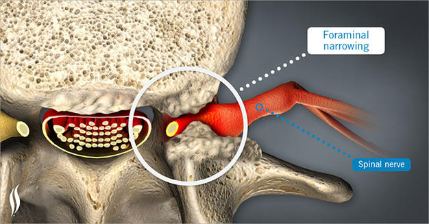
Fig. 2: A lumbar nerve root is seen pinched between arthritis as it emerges through the neuroforamen. This condition is relatively common and will not respond well to conservative management. Reducing lumbopelvic arching (only applicable to this specific condition), repeated nerve root block procedures, and taking it easy can help, but only surgical liberation of the foramen will cure the problem.
The next condition that I frequently come across, being totally misunderstood, are smaller lesions in the co-presence of a small spinal canal. The width of the spinal canal is determined by the length of the pedicles, and angle of the laminae. Generally, one would expect a large hernia or a lot of arthritis to be able to cause chronic nerve compression in the spine. However, in cases where the canal is small, a smaller or even much smaller lesion is required to create nerve compression. I have seen many cases where a virtually normal spine, perhaps just 1mm bulge (which is nothing!) of a disc is enough to cause chronic compression of certain nerves due the narrow environment for the nerves that is caused by the short pedicles (small canal). These cases are almost always reported as normal, yet will not heal spontaneously! For accurate prognostic evaluation, an MRI, and understanding of this phenomenon is absolutely warranted.
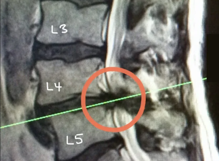
Fig. 3: Although this is a red flag, it gets the point across. A patient born with a very small spinal canal has developed a relatively small herniation and has a little posterior arthritis and thickening of the ligamentum flavum. This, in this case, was enough to shut the entire canal! A patient with a normal canal caliber would usually not even see mild nerve compression with a same-sized hernia.
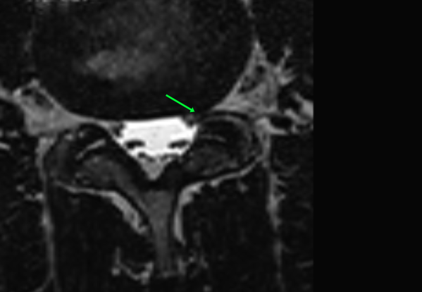
Fig. 4: Here is an interesting case, where a patient with a virtually normal spine had the ever-so-slightest bulge of the L4-5 disc. Only due to his short pedicles, the minuscule bulging of the disc was enough to cause chronic compression of the left L5 nerve at its descending path between the disc and the lamina. This patient had 5 different reports who all stated his findings were normal. He ended up paying an out-of-pocket surgery where they scraped open the lamina in the back to liberate the nerve, and the issue resolved.
Disc thinning and Modic changes
Finally, the final commonly misunderstood problem pertains to disc thinning and related Modic changes. This is usually reported as “age-related wear and tear” and tends to be discarded as non-important findings. It is true that some patients have this and are asymptomatic, so you always want to look for compatible symptoms and triggers before doing anything rash. However, many patients with disc thinning and Modic changes have severe symptoms.
Modic changes, although somewhat controversial, occur when the disc has lost its normal shock-absorbtive capability, causing bending and impacts to irritate the hard spine rather than being sucked up by the soft discs. Over time, this causes end plate changes in the vertebrae and can lead to severe inflammation of the bone in some cases. It is, or at least starts as, osteoedema; not a mystery. It can later sclerose, which is why it becomes hypointense on T2 imaging and hyperintense on T1 (for the interested reader). In rare cases, it can become infected and will require antibiotic treatment over longer periods of time, often intravenously.
Patients with symptomatic Modic lesions (presuming there are no other issues) will feel excruciating pain deep inside the spine, and it will typically not disseminate to other areas. These patients will improve with rest and grossly worsen with bending and movement of the spine. When the findings are mild, significant improvement can be seen with conservative management. However, when the disc height is so reduced that abnormal shock of the vertebrae is unavoidable, then installing a disc prosthesis is a good idea that can be totally curative if the diagnosis stilled,- and the surgical procedure are both done properly.
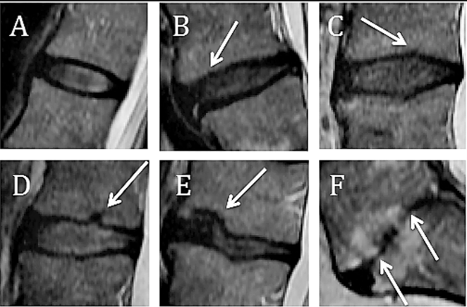
Fig. 5: Here we see degrees of disc detriment. Somewhere between E and F, or even at E if sufficiently symptomatic, then a disc prosthesis could be contemplated. A-D can all be treated conservatively with good results. Once at F, where the disc is totally destroyed and the end plates have severe damage, then installing a prosthesis is often rendered impossible. In these cases you will have to wait for natural fusion, which can take years. I discourage surgical fusion of the low back unless absolutely warranted. Although surgical fusion tends to work well for the neck, I have seen mostly poor outcomes pertaining to the lower back.
Sacroiliac joint pain
This will just be a short section. Sacroiliac joint (SIJ) pain is a controversial issue with much misleading information around it. Some claim it doesn’t even exist, whereas others claim it is responsible for a huge proportion of low back problems (Vleeming, 2012; Dontigny, 1990). I have found this condition to be rare, and that most patients with SIJ-area symptoms actually suffer from middle and superior cluneal nerve symptoms. I will get back to this later, in brevity.
Patients with genuine SIJ pain will feel a sharp, very distinct, very focal (exact location) pain inside of the sacroiliac joint, usually on one side, and usually when bending, sidebending or rotating. They do NOT tend to get worse when sitting; this is the middle cluneal nerve (a mimic, explained later). The pain will typically NOT spread, unless the extreme rarity of a sacroiliac upslip (which we are not getting into here). When it is really bad, even when walking. Most patients who get this are hypermobile athletes or athletes whose sport is extremely monotonous in terms of movement patterns, eg. javelin throwers, where they almost always rotate the same way, all the time. Patients with autoimmune disease are also more prone to develop autoimmune sacroiliac joint inflammation, which is frankly more common than genuine mechanical sacroiliac joint dysfunction in my experience.
There are various physical tests that can be done for this condition, but ultimately, its cause is either ligamentous damage and/or severe hip muscle function imbalance. I have found that the most important muscles to rehabilitate in order to improve genuine mechanical sacroiliac joint dysfunction are the piriformis (most important one), psoas major, quadratus lumborum and multifidus muscles. All of the aforementioned can be found on my youtube channel. Train no more than twice per week.
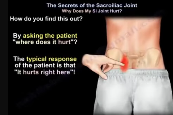
Fig. 6: Fortin’s finger test: Patient points exactly to the SI joint fossa every single time they are asked to point. Must be replicated at least three times. Pointing out different places would suggest another issue than genuine SIJD.
Myogenic low back pain
Statistically, and practically, the most common source of muscular low back pain is the quadratus lumborum muscle. The quadratus lumborum is situated in a vulnerable position because it is highly stretched out when you bend forward or sidebend, making it prone to tearing if it is weak and overburdened. Patients complaining of quadratus lumborum pain will typically have recurrent bouts of pain that come after repetitive bending and loading, especially squatting and deadlifting in the gym. This can affect old as well as young patients. The pain will usually be situated at the posterior flank, usually one side but can be both. When ongoing, it will typically trigger with sidebending to the opposite side, or bending forward with sidebending to the opposite side, as this stretches the muscle. In clinic, I push my thumb hard into the QL and look for reproduction of the same symptoms.
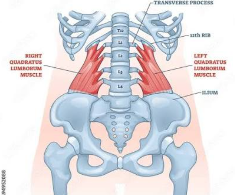
Fig. 7: The quadratus lumborum muscles attach between the 12th rib, transverse processes of the lumbar spine, and iliac crests. It works well when strong, but if weak, which is common, is highly prone to tearing when stretched under load.
The reason why the quadratus lumborum becomes weak, is usually postural. Most patients have an inadequate lumbar lordosis in posture, and this reduces or even inhibits the quadratus lumborum, leaving it to wither in many cases. When a certain degree of inadequacy is reached, it will become prone to strain and tearing, often recurrent. Quadratus lumborum injuries can cause debilitating pain, but is generally self-resolving. The issue is that it keeps occurring at gradually greater frequency until the underlying issue is resolved.
Stretching, massages and needling will all make it worse, even if you feel better in the short term. For the love of God, do not do botox; this will often decay the muscle to such an extent that recovery is very difficult. Gentle strengthening of the muscle done twice weekly is what is required to improve. If you train and feel worse, then do less. The aim is mild symptom worsening for 1-2 days per week. You should keep doing the exercises for at least a full year after the pain is gone to prevent recurrence.
Injuries to the erector spinae are rare, but would be treated the same way, but with a different exercise, eg. the birddog. I have various exercises on my youtube channel. The psoas major is another common culprit, but because it tends to cause predominant nerve symptoms, it comes under the next subchapter.
Neurogenic low back pain / Nerve entrapment syndromes
Despite being a mysterious etiology of low back pain, nerve entrapment syndromes affecting the low back are a very common problem (Larsen, 2019). The medical literature has sparse information on this topic, and most of these patients will ultimately be diagnosed with fibromyalgia if they complain sufficiently to their primary care provider. I suggest that you read my article on lumbosacral plexus entrapment syndrome for elaborated information on this topic, but I will write down the essential information here.
The lumbar and sacral nerve centers, also known as the lumbar and sacral plexuses, both emerge through tight compartments that can be compromised. The majority of the lumbar plexus emerges through the fibers of the psoas major, whereas the sacral plexus emerges between the superior gemellus and piriformis muscles. Severe weakness of these muscles is common, and at a certain level of detriment, consequent scar tissue formation can cause the nerves to develop friction in their neurofascial gliding canals, leading to nerve pain. This can range from very mild to very severe. Unfortunately, most patients will, either by being told to do so or by doing their own research, end up doing stretches for these muscles. Similarly to the QL, it can feel helpful in the beginning, but will sadly result in worsening over time. Weak muscles get worse from releasing, and this is an important fact to be aware of!
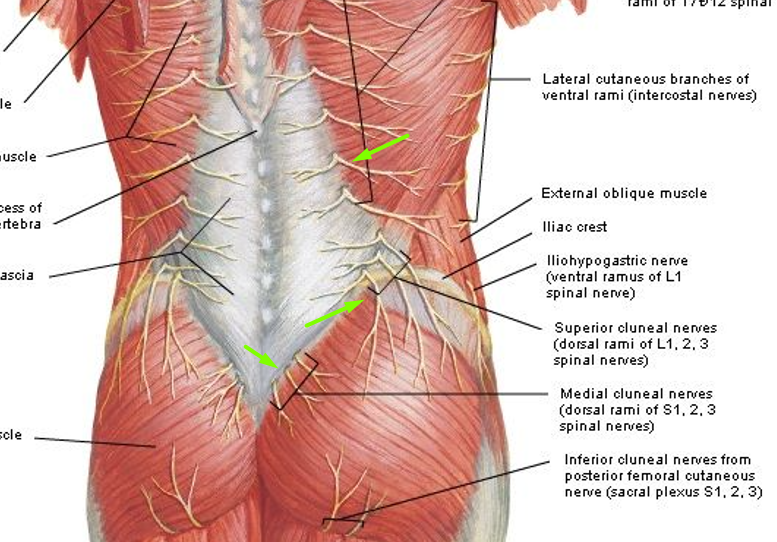
Fig. 8: Note how the middle cluneal nerves differ from the superior nerves both in location, but most importantly, stemming from the sacral rather than lumbar plexus. Entrapment of the middle cluneal nerves is the most common cause of symptoms in the sacroiliac area, and is a mimic. Palpating the area where they emerge will be abnormally painful. The same applies for the superior cluneal or dorsal rami nerves, which stem from the lumbar plexus.
Because this article is about low back pain, there are two main nerve zones that will be relevant; the dorsal rami nerves of the lumbar spine, and the middle and superior cluneal nerves, as briefly mentioned earlier. The superior cluneal nerve and dorsal rami nerves are both emanating from the lumbar plexus, and thus, carefully rehabilitating the psoas major muscle is the appropriate treatment for this condition. On the other hand, the middle cluneal nerve, which is the most common cause of sacroiliac area pain and misleading mimic of SIJD, leading to misdiagnosis, emanates from the sacral plexus, and thus, the appropriate treatment for this condition is directed to the piriformis and deep six hip rotators.
For the dorsal rami nerves, pain tends to be felt a couple of inches lateral to the spine, often at the edge of the iliocostalis muscle. Palpating this area will be abnormally painful, and the patient will have difficulties pinpointing the exact location of the pain (this goes for most of these nerve entrapment conditions). For the superior cluneal nerve, the pain is located at the flank of the buttock, approximately from mid-glute (in the gluteus medius area) and going up to the iliac crest. Finally, but as already explained, the middle cluneal nerves tend to cause pain around the sacrum and SI joints, it is often sided but can also be bilateral. Patients with middle cluneal neuralgia tend to worsen when they sit, which genuine mechanical sacroiliac patients do not. For the superior cluneal and dorsal rami symptoms, worsening is typically seen after activity that loads the hip flexors, such as walking and running, especially uphill.
As with the quadratus lumborum, strengthening is done very carefully twice per week. Find a low repetition amount that yields mild worsening the next day, and do that twice per week. Any more than mild, or any more than 1 day of worsening (max. two days per week) will prevent improvement, and can even cause worsening, so approach this with care.
Clenching issues
I shall, finally, reconcile my statements with some biopsychosocial consensuses. And, although it comes last, it is not less important than the aforementioned, and is especially pertinent for patients with chronic pain. Most patients with chronic musculoskeletal maladies that do not stem from physical trauma (eg., your back started hurting after you got run over by a tractor) have tendencies for clenching. This means that the patient has a tendency to disproportionately clench / overactivate certain muscle groups, which leads to excessive and refractory loads on the lower back, even when they are at rest. I call this phenomenon clenching and bracing patterns (you should read this article (link)).
For low back pain, the most common “clenches” that is seen in average patients with longstanding issues, are buttock and abdominal clenching, without a doubt. Clenching of the buttock and abdomen, one or the other, both result in flexion of the lower back. Constant flexion of the lower back is not only bad for the discs, as showed earlier, but also completely disengages the low back erector musculature! In other words, not only does it place you in a constantly unfortunate angle for your lumbosacral spine, but it also takes away the spine’s local support! To top it off, constant clenching, and probably abdominal clenching more so than buttock clenching, will also increase the load on your spine.
So, in addition to improving your posture as I have explained earlier, you must also ascertain that the abdomen and buttocks are not being clenched in posture. I go into this briefly in some of the videos in the beginning of this article. Furthermore, a couple of videos with elaborated information will be added here.
SOURCES:
- Engelmann P, Busmann M, Löwe B, Hüsing P. Biopsychosocial risk factors for the transition from acute to chronic back pain: A prospective cohort study. J Psychosom Res. 2026 Mar;202:112500. doi: 10.1016/j.jpsychores.2025.112500. Epub 2025 Dec 13. PMID: 41500080.
- J Bodyw Mov Ther. 2011 Apr;15(2):131-8. doi: 10.1016/j.jbmt.2011.01.011. The fall of the postural-structural-biomechanical model in manual and physical therapies: exemplified by lower back pain. Lederman E1.
- Chronic diseases and health promotion: http://www.who.int/chp/topics/rheumatic/en/
- Keith L. Moore, 2007, «Essential clinical anatomy, 3rd)
- Marshall LW, McGill SM. The role of axial torque in disc herniation. Clin Biomech (Bristol, Avon). 2010 Jan;25(1):6-9. doi: 10.1016/j.clinbiomech.2009.09.003.
- Veres SP, Robertson PA, Broom ND. The morphology of acute disc herniation: a clinically relevant model defining the role of flexion. Spine (Phila Pa 1976). 2009 Oct 1;34(21):2288-96. doi: 10.1097/BRS.0b013e3181a49d7e.
- McGill SM, Hughson RL, Parks K. Changes in lumbar lordosis modify the role of the extensor muscles. Clin Biomech (Bristol, Avon). 2000 Dec;15(10):777-80.
- Aultman CD, Scannell J, McGill SM. The direction of progressive herniation in porcine spine motion segments is influenced by the orientation of the bending axis. Clin Biomech (Bristol, Avon). 2005 Feb;20(2):126-9.
- Tampier C, Drake JD, Callaghan JP, McGill SM. Progressive disc herniation: an investigation of the mechanism using radiologic, histochemical, and microscopic dissection techniques on a porcine model. Spine (Phila Pa 1976). 2007 Dec 1;32(25):2869-74. doi: 10.1097/BRS.0b013e31815b64f5.
- Wade KR, Robertson PA, Thambyah A, Broom ND. How healthy discs herniate: a biomechanical and microstructural study investigating the combined effects of compression rate and flexion. Spine (Phila Pa 1976). 2014 Jun 1;39(13):1018-28. doi: 10.1097/BRS.0000000000000262.
- Potvin JR, McGill SM, Norman RW. Trunk muscle and lumbar ligament contributions to dynamic lifts with varying degrees of trunk flexion. Spine (Phila Pa 1976). 1991 Sep;16(9):1099-107.
- Dangaria TR, Naesh O. Changes in cross-sectional area of psoas major muscle in unilateral sciatica caused by disc herniation. Spine (Phila Pa 1976). 1998 Apr 15;23(8):928-31.
- Barker KL, Shamley DR, Jackson D. Changes in the cross-sectional area of multifidus and psoas in patients with unilateral back pain: the relationship to pain and disability. Spine (Phila Pa 1976). 2004 Nov 15;29(22):E515-9.
- Freeman MD, Woodham MA, Woodham AW. The role of the lumbar multifidus in chronic low back pain: a review. PM R. 2010 Feb;2(2):142-6. doi: 10.1016/j.pmrj.2009.11.006.
- Kader DF, Wardlaw D, Smith FW. Correlation between the MRI changes in the lumbar multifidus muscles and leg pain. Clin Radiol. 2000 Feb;55(2):145-9.
- Cooper RG1, St Clair Forbes W, Jayson MI. Radiographic demonstration of paraspinal muscle wasting in patients with chronic low back pain. Br J Rheumatol. 1992 Jun;31(6):389-94.
- Parkkola R, Rytökoski U, Kormano M. Magnetic resonance imaging of the discs and trunk muscles in patients with chronic low back pain and healthy control subjects. Spine (Phila Pa 1976). 1993 Jun 1;18(7):830-6.
- Landmark L, Sunde HF, Fors EA, Kennair LEO, Sayadian A, Backelin C, Reme SE. Associations between pain intensity, psychosocial factors, and pain-related disability in 4285 patients with chronic pain. Sci Rep. 2024 Jun 12;14(1):13477. doi: 10.1038/s41598-024-64059-8. PMID: 38866885; PMCID: PMC11169509.
- Larsen K. Common low back pain, is it really a mystery? Anaesth Pain & Intensive Care 2018;22(1):125-130
- J Anat. 2012 Dec; 221(6): 537–567. Published online 2012 Sep 19. doi: 10.1111/j.1469-7580.2012.01564.x PMCID: PMC3512279 The sacroiliac joint: an overview of its anatomy, function and potential clinical implications A Vleeming,1,2 M D Schuenke,1 A T Masi,3 J E Carreiro,1 L Danneels,2 and F H Willard1
- DonTigny RL. Anterior dysfunction of the sacroiliac joint as a major factor in the etiology of idiopathic low back pain syndrome. Phys Ther. 1990 Apr;70(4):250-65; discussion 262-5. doi: 10.1093/ptj/70.4.250. PMID: 2138334.
- Larsen K, Chien GCC. Lumbosacral plexus entrapment syndrome. Part one: A common yet little-known cause of chronic pelvic and lower extremity pain. Anaesth pain & intensiv care 2019;23(2):124-137
