
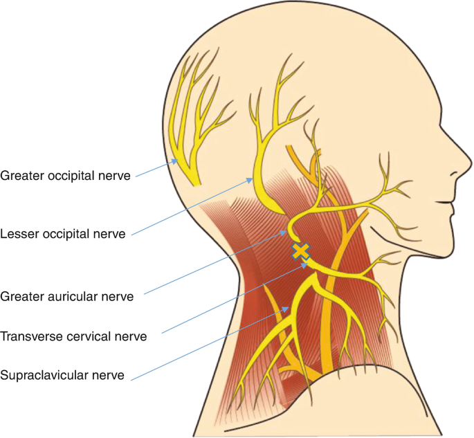
The Cervical Plexus Entrapment Syndrome (CPES) is a condition that you will find little to no information about online, but is still a relatively common disorder in the chronically ill, and is something I have been researching over the last decade of clinical practice. It is not particularly common in the general population. There are a few papers presenting cases where upper cervical radiculopathy has resulted in a cervical plexus syndrome (Kessler 1991), and of course, injuries to the plexus through traction and nerve avulsion, nerve tumors (Gierek 2003) or deemed idiopathic (Uterga 1998). What we are talking about here, however, is an entrapment syndrome of the cervical plexus, and to the best of my knowledge, I have not seen anyone else mention this problem nor present a potential pathomechanism. I first mentioned it in one of the first exhaustive neck pain articles that I wrote on this website in 2017 (link).
Pain and paresthesia in the face is poorly understood, but is generally expected to be stemming from the sinuses, teeth, temporomandibular joint, or intracranial lesions (LESSE 1956). When neither of these can be proven, the symptoms are often deemed idiopathic, or psychogenic (Naidu 2020). The cervical plexus is rarely considered as a potential cause for the patient’s symptoms due to the very sparse medical literature available on the topic. Moreover, as with other entrapment plexopathies, the symptoms vary from patient to patient, which makes it hard for the examining clinicians to establish a clear pattern. This, clearly, also makes it much harder to produce pragmatic and practical papers on the topic.
This is an article I’d been meaning to write for years, along with the Longus capitis syndrome, which is up next. I was initially planning on publishing this academically, and I still am, but the large/formal writing projects are not coming out as I had hoped. So, this will have to do for now.
Cervical plexus anatomy
The cervical plexus forms from the C1-4, and partially the C5 nerve roots. It provides sensory innervation around the neck, shoulders and face, making it overlap with other potential problems. This can be very confusing when diagnosing it, and it is crucial to understand where the symptoms may present as well as what the expected triggers are.
The cervical plexus carries various muscular branches, the most important of which being the 3rd to 5th roots, which innervate the diaphragm. The ansa cervicalis is formed between the C1-3 nerve roots, and innervate all of the strap muscles of the throat, ie. the infrahyoid- and suprahyoid muscle complexes, which are involved in speech, swallowing and various jaw movements. The C1-3 nerve roots also innervate the suboccipital muscle group, rectus capitis lateralis & rectus capitis anterior, and the longus capitis — ie. the craniocervcial stabilizers. Finally, it contributes to some of the paraspinal erectors, the sternocleidomastoideus & trapezius muscles (via the spinal accessory nerve), as well as the scalene and levator scapula muscles.
The motor function of these nerves is generally not very important in a setting of cervical plexus entrapment syndrome, because myofascial friction is typically not hard or rough enough to produce sinister or considerable nerve damage (weakness). Instead, friction and irritation of the myelin sheath around the nerves results in pain, but not weakness. Therefore, if there is blatant weakness of any of these motor pathways, CPES is unlikely to be the cause. A brain and spine MRI should be done instead, or even workups for systemic causes if the clinical picture suggests that.
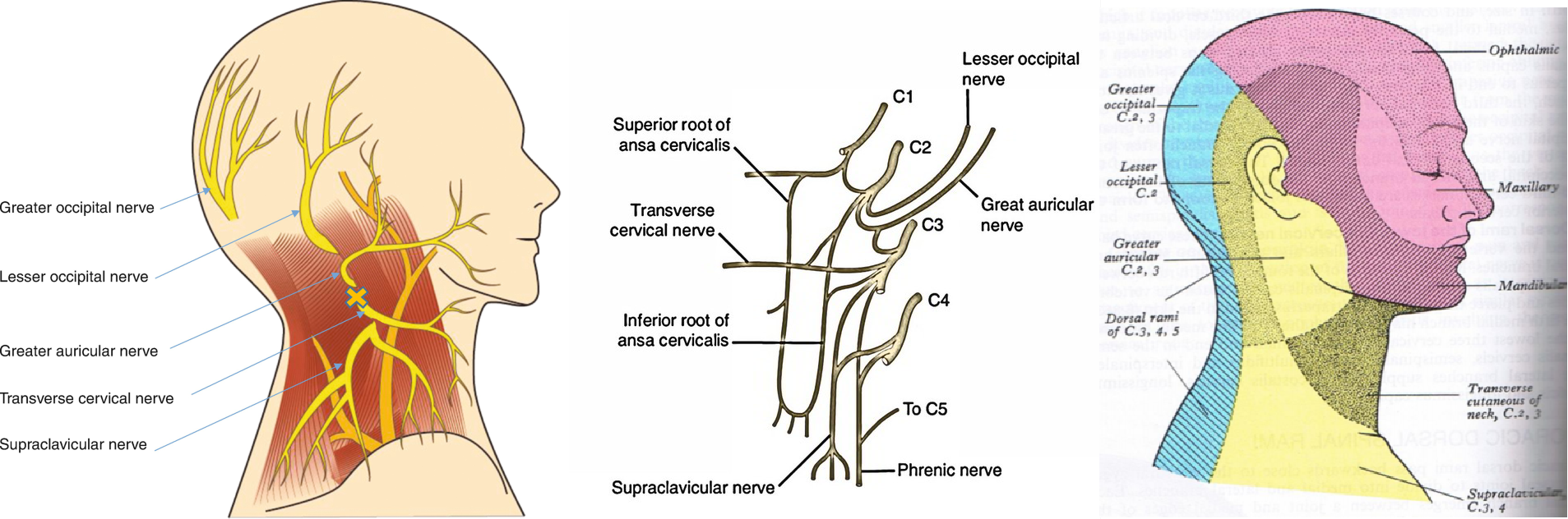
As shown above, the main symptom of nerve affection in cervical plexus entrapment syndrome is pain and paresthesia, not weakness. It is therefore important to be familiar with its zones of sensory innervation. You may have heard about occipital or auricular neuralgias before. These conditions can be caused by focal entrapment as a single nerve entrapment, or be secondary to entrapment at a plexus site. However, the reach of the cervical plexus exceeds the mere back of the head. Through its dorsal rami, symptoms can arise along the upper back, especially close to the spine, such as between the shoulder blades. It can also influence the dorsal scapular nerve, which is a periscapular nerve. It influences the area around the ear through its auricular nerves, and below the jaw through the transverse cutaneous nerve. Finally, the supraclavicular nerve innervates the anterior cutaneous aspect of the trapezius, including the supraclavicular skin. As you can see, these areas overlap with other conditions, such as thoracic outlet syndrome, TMD and Costen’s syndrome, cervical radiculopathy, mononeuropathy (eg. occipital neuralgia), and more, making a conclusive diagnosis difficult without the right diagnostic approach.
The ansa cervicalis nerve anastomoses with the hypoglossal nerve (cranial nerve XII), and it is theoretically possible that derangement of the cervical plexus would also, then, be able to cause symptoms in the tongue and other structures that are innervated by the hypoglossal nerve. In my experience, however, this is not common, and I would only consider it if I was able to trigger a potential tongue symptom along with other “blatant” cervical plexus symptoms, and of course, triggered by one of the entrapping culprits for CPES, which we will review later.
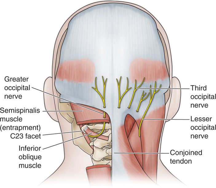
The cervical plexus emerges between the anterior and middle scalene, before continuing between the levator scapula and sternocleidomastoideus muscles. It can be entrapped by any of these structures, but for CPES, the levator scapula is the most common instigator. Further, the occipital nerves, although not the main topic of this article, can become entrapped within the tissue of the upper trapezius and semispinalis muscles, as they pierce them. In many cases of occipital neuralgia, the nerve may be pre-irritated at the plexus, then adding a “double crush” phenomenon at the trapezius, which is ultimately what instigates the symptom. Therefore, even in mononeuropathies, it is useful to be aware of the nerve’s course from the root, all the way to the respective end point. You will want to make note of which tissues it passes intimately adjacent to, or through.
Symptoms & diagnosis
Having knowledge about the mere existence of cervical plexus entrapment syndrome is definitely the first and most important step in the diagnostic process. How can you diagnose something you do not know exists? Very difficult, which is why it took me several years to map the information that I am providing in this article. Now, the next step, as stated, is to be familiar with the potential symptoms that can manifest in this condition, and as importantly, its relevant triggers. In essence, true CPES is a condition that is triggered by provocation of its entrapping muscle(s), and from experience I can assure you that the most common cause is the levator scapula muscle, even though the sternocleidomastoideus and scalenes may also be implicated.
The symptoms in question are usually either pain or paresthesia, without neurogenic weakness. The locations of the symptoms are common along the entire reach of the cervical plexus, and thus I cannot give you a single “most common” location, as I would with TOS (which would be between the shoulder blades and along the ulnar nerve). The symptoms usually feel superficial, not deep in the tissues. A patient afflicted by cervical plexus entrapment syndrome will typically state that they have pain or discomfort in the face (but no higher than the cheek-bone, this is important), around the ear, below the jaw (in the submandibular area) or back of the head, if we’re talking about the head region. In the neck, the most common areas are the front and side of the neck, but can sometimes also manifest behind the neck. Because the 5th cervical nerve partially emanates from the cervical plexus, you may also see symptoms in the scapula (suprascapular nerve), between the shoulder blades (dorsal scapular nerve), and even the shoulder itself (axillary nerve). Symptoms in the arm are not expected in a case of sole cervical plexus affliction.
Symptoms between various conditions can greatly overlap, and therefore, having compatible symptoms is only enough to raise suspicion, but not sufficient for a conclusive diagnosis. You will, preferably, also find compatibility with regard to history as well as triggers.
Developing CPES secondary to some kind of head/neck injury is definitely the most common, because it can injure the very muscles that entrap the plexus. Shoulder and collarbone injuries can also damage the levator scapulae. Egregious, long-term stress can cause it as well, but I will not be discussing the stress mechanism in this article. A CPES patient will often tend to say that their symptoms get better when they avoid moving or loading their necks too much. They will frequently have limited range of motion, and can feel cracking and popping from the cervical facet joints, especially in the upper parts of the neck. The levator scapula instigates ipsilateral rotation of the neck, and therefore stretches with contralateral rotation. This means that if the patient has restricted rotation to the right, for example, but feels the restriction on the opposite side, then that suggests an issue with the levator scapula. If the restriction and symptom is on the same side, then this would often suggest a scalene or trapezius problem, but beware that these can occur simultaneously due to the mechanism of injury.
Palpation of the levator scapula will elicit abnormal amounts of pain in almost all of these cases. However, in a case of whiplash, many cervical muscles can be affected. Do not let this trick you into believing that the patient is “sensitized”; this will not help anyone. If the patient presents with pain within the reach of the cervical plexus, has a history of some kind of head or neck injury, or long-term severe stress, has restricted rotational range of of motion and/or upper cervical crepitus, and also demonstrates severe palpation tenderness along the levator scapula and sternocleidomastoideus muscles, then cervical plexus entrapment is a realistic potential explanation for this individual.
An Erb’s point block can be done, but I have found it unreliable as an absolute diagnostic marker. If an Erb’s point block is done, and ameliorates the pain for a few hours, then this can be considered a diagnostic confirmation. However, if it does not, then this would not disprove the presence of CPES for a patient who carries other clear symptoms of such. Also, for greater occipital neuralgia, injection may also be required at its passage just lateral to the occipital protuberance.
The final step is to perform specific provocation for the very culprit muscles, and see if you can reproduce the symptoms. I will typically start with the levator scapula for this, as it is the most prevalent offender and also more specific to this condition. I recommend gentle levator scapula strengthening done once to twice per week, typically only one work-set per session. Look for worsening of symptoms the next day, although it can sometimes take longer. If this happens consistently, then you have found the offender and have confirmed the diagnosis. At this point, I usually implement the scalene and sternocleidomastoideus muscles as well.
The reason why I do not recommend starting with the scalene and sternocleidomastoideus muscles, is that they can influence other conditions as well. This is not helpful during the initial diagnostic work, but it should certainly be done after the diagnosis has been established. Because of the intimate anatomical relationship with the scalenes and the levator scapulae, one could see how both symptoms and triggers can be easily mistaken for thoracic outlet syndrome, but get confused due to incompatibility between upper cervical and facial symptoms and the reach of the brachial plexus.
In summary:
- Pain or paresthesia within the reach of the cervical plexus
- History of head/neck injury or severe emotional distress
- Restricted cervical range of motion
- Popping or cracking in the C1-4 facet joints
- Massive pain upon palpation of the levator scapula
- Reproduction of symptoms after systematic exercise-induced provocation of the levator scapula
- Negative MRI
- Typically, no one else has been able to “find anything” concrete, despite the patient having visited a plethora of clinicians
Imaging and electrodiagnostic studies
CPES is not a condition that is confirmed by imaging nor electrodiagnostic workups. Imaging will also not tend to reveal entrapment of plexus in soft tissue, as has been well documented in N-TOS research. A systematic review by Kwee et al. (2014) showed that magnetic resonance neuropraphy was an unreliable assessment in nerve entrapment disorders in general (Kwee 2014). Moreover, electrodiagnostic exams will in almost all cases be negative when there is no blatant neurogenic weakness. Conditions that are prone to genuine nerve injury, eg. carpal tunnel syndrome, are much more detectable through neurographic studies. This has led many to believe that most other entrapment sites are mere myths and legends without holding in real science (eg. piriformis syndrome), and causes many of these patients to be stigmatized and misdiagnosed with psychosomatization or a fibromyalgia. A paper published by Rousseff et al. (2005) showed that 80% of patients with surgically proven thoracic outlet syndrome had completely normal electrodiagnostic tests (Rousseff 2005). Keep well in mind that TOS leads to genuine weakness of the arm quite frequently, due to hard compression (as opposed to myofascial friction) of the brachial plexus in the costoclavicular space. Therefore, one may appreciate how conditions that only involve myofascial irritation (which does not tend to lead to nerve weakness due to insufficient compression of the nerve) almost never demonstrate concrete, reliable abnormalities upon electrodiagnostic testing.
Thus, understand that a normal MR neurography, EMG, NCV or cervical spine MRI does not exclude the presence of CPES.
Differential diagnosis
A full review of differential diagnoses and their signs and symptoms is outside of the scope of this article. However, you need to be aware that various unrelated clinical entities can cause similar symptoms. Which conditions need to be excluded would depend on where the symptoms are manifesting, and which triggers the patient has.
- Facial symptoms: Exclude temporomandibular disorder as well as intracranial lesions that could affect the trigeminal and facial nerves. In a case of TMD, chewing, talking and jaw clenching would all be expected to trigger their symptoms. In a case of intracranial impingement of the nerves, however, the symptoms would typically arise “out of the blue”. I generally acquire a brain MRI for patients with facial symptoms, just in case.
- Neck pain: Exclude cervical radiculopathy and thoracic outlet syndrome. The first mentioned is excluded by means of a non-contrasted cervical spine MRI, but TOS is trickier. TOS and CPES often co-exist. Its diagnostic workup exceeds the scope of this article. Beware that brachial plexus impingement can mimic CPES, and often trigger with arm-loading due to subsequent narrowing of the costoclavicular space. This is a common clinical dilemma, and if I suspect CPES vs. TOS I tend to just commence treatment for both.
- Shoulder and shoulder blade: Exclude cervical radiculopathy. Exclude TOS.
- Diffuse pain without a history that would explain them #1: Consider excluding systemic causes, such as small fibre neuropathy. If your client has a history, or family-history of diabetes or autoimmune diseases, then having your patient examined by a rheumatologist could be necessary as reassurance.
Exclusion of other potential diagnoses can be a time-consuming and costly endeavor that may be essential for some patients, but much less so for others. For example, if a person underwent a neck-whipping incident and now suffers from CPES-compatible symptoms, never having any other history of such, then proceeding with the clinical diagnostic process is a viable option. Especially if the symptoms had onset not too long ago. You could do a cervical spine MRI, just to be thorough. If the clinical workup reproduces his or her symptoms, then commencing treatment is reasonable. However, if the same symptoms occur “out of the blue”, and the patient denies any head/neck injuries, sports injuries, or severe stress in the period leading up to the onset of their symptoms, then care must be taken to diligently exclude other potential causes. Especially those with potentially sinister consequences, such as an intracranial lesion, MS or other important condition.
Treatment
When determining the treatment strategy for CPES, it is always helpful if one has a grasp of the underlying cause. Especially with regard to factors that may still be present.
If a patient develops CPES after a neck injury, then that incident was the cause. If you diagnose and treat the consequences (ie. the muscular injuries) properly, the patient will improve. However, if the individual has developed CPES or similar symptoms secondary to severe anxiety (and neck clenching), for example, then both conditions need to be treated simultaneously. Treatment of the symptom will be futile, or at best ineffective, if the cause is still there.
The levator scapula is always involved, at least as far as I have seen. The levator scapula is an important stabilizer of the shoulder blades, especially during horizontal and overhead loading. It is also an important stabilizer of the upper cervical spine. I generally start with management of the levator scapula, along with correctives for shoulder posture and scapular dyskinesia, if applicable. Even in cases where scapular movement is normal, the patient may be causing overloading of the levator scapula muscle by various activities that they are involved in, and this might have to be reduced for a period of time.
- Try to determine whether the patient’s CPES was induced by a mechanical injury (which is most common), stress (less common), or a shoulder/collarbone injury (least common). Patients with stress-induced CPES need to learn how to stop neck clenching (link).
- Do the patient’s triggers suggest that neck loading alone, or also shoulder-loading is triggering their symptoms?
- Start careful strengthening of the levator scapula, scalenes and sternocleidomastoideus muscles. This should be done with great care, once to twice weekly, at one single work-set to moderate fatigue.
- If the patient is also triggered by shoulder loading, then additional work for scapular correctives may be required. This can easily be overdone and lead to overloading of the levator, so implementation must be done carefully, and be monitored.
- In cases where stress is culpable, it is usually because the patient neck “guards” / clenches in response to that emotional distress. This guarding technique can lead to tremendous overwork of the neck. This can be identified and mitigated, as explained in the article that I linked above.
- Avoid stretching, deep/aggressive tissue massage, dry needling, and botox. Botox is the worst of all of them. All of the above will worsen the situation, even if you may feel some initial improvement. I am basing this statement on hundreds of these cases.
The levator scapula muscle can be very difficult to engage, even with specialized exercises. It is the most important factor of CPES management. The levator is vulnerable to stretch injuries during whiplash due to its long span between the superior angle of the scapula, and the upper neck. It is vilified for being “overactive” and a culprit in, a notion I subscribed to for many years. Eventually, I realized that the levator scapula was just another victim, and required strengthening. Further proof is easily obtained when even the slightest stimulation of the levator, in these cases, leads to worsening. If the levator scapula was truly “overactive” and “strong”, would you not see a great capacity to load? This is not the case, and I strongly recommend that you heed my warnings.
This muscle has many roles
- Extension of the neck (contraction)
- Controlled flexion of the neck (lengthening)
- Ipsilateral rotation of the neck
- Stabilization of the upper cervical spine
- Downward rotation of the scapula
- Protraction of the scapula
- Anterior tilting of the scapula
- Oblique elevation of the scapula during anterior tilting
- Preventing excessive upward rotation and posterior tilting of the scapula
I have found that treating the levator scapula as an “elevator” will generally result in failure. The levator scapula is much more active in horizontal stabilization than in the vertical sense. Prepping the scapula by pulling your arms back will tilt the scapula into anterior and downward tilting. From this position, lengthen and slightly protract the neck. Finally, maintain that position as you very slowly pull the scapula forward towards the chin. The movement should be forward and ever so slightly upward, NOT downward, as the latter would engage the pectoralis minor. Unfortunately, this is an intricate procedure, and I have seen highly intelligent and motivated patients fail for long periods of time. But, it is possible with the right technique.
Next up are the scalene and sternocleidomastoideus muscles. The scalenes perform lateral flexion of the neck, whereas the sternocleidomastoideus causes contralateral rotation and flexion of the neck. Ie., their rotational purposes are opposite, and the flexion trajectories are also different from one another. The videos are old, so try to accommodate what I write in the text while only using the videos as a rough reference. Beware, however, that performing these exercises correctly makes you much more prone to severe post-exercise inflammation if you pertain to these patient groups (including TOS as well). Please be careful. Careful explanations of scalene and SCM training can be found in my TOS (link) and upper cervical (link) articles.
For the scalene, lie on your side with your torso reclined to approximately 70 degrees (presuming lying straight on your side is 90). Draw your head slightly forward, then rotate your head toward the ceiling. Keep your chin up, and aim with your chin/nose, then pull your head slowly towards the ceiling. Do not aim for the shoulder; aim for the ceiling.
For the sternocleidomastoideus, you will be situated on your back. Turn your head to one side, but not toward the shoulder; keep the head centered between your two shoulders. Keep the chin up slightly, and aim with the ear/back of your head. Draw your ear slowly towards your sternum without losing the contralateral rotation.
As with all of these exercises, careful implementation is key. A patient with CPES will be highly sensitive to overtraining, and this will occur in a much different way than conventionally expected. Many, indeed, most patients and clinicians alike expect overtraining to occur after brutal and obvious excess. Please understand that this does NOT apply to these patient groups: For an individual where mere movement of the neck triggers their symptoms, overtraining comes extremely quickly. You must beware of this in advance, and make the necessary preparations. So many patients attempt strengthening, but proceed to get worse, then discouraged, and give up, and go back to massages and stretches. This will never resolve your problems. The reason you get worse, is that the muscles have inadequate work capacity and get overly inflamed from the work you have imposed on them. Much less work is required in order to see progress. And, of course, patience. I generally recommend one single work-set done once to twice weekly, for each muscle. Aim for one single day of mild worsening after training; this is the sweet spot. If you can get 1-2 days of very mild worsening after training, consistently, then progress will be seen. On the same coin, those who do not respect these crucial signs/limits, will inevitably fail. I have sadly seen this happen countless times.
Scapular correction can be very complicated, and is also not necessary in all CPES patients. The intricacies of scapular movement corrections exceed this article, but are elaborated to a great extent, and for free, in this article (link). I will, however, add a video below. If shoulder use triggers your symptoms, keep it to a minimum until you have gotten further in your neck rehabilitation. Expect at least six months, often longer if there is greater severity.
Neck posture, although important in a general sense, is not a key player in CPES management. Most people have suboptimal neck postures, and still have no signs of CPES. However, exceptions may be important in individual cases. What is of crucial importance, however, is the abolishment of any neck clenching habits. For your leisure, I attach some howtos below.
Finally, I will adhere a video, for your leisure, on the basics of clenching assessments.
References
- Kessler LA, Abla A. Syndrome of the cervical plexus caused by high cervical nerve root compression. Neurosurgery. 1991 Apr;28(4):506-9. doi: 10.1097/00006123-199104000-00004. PMID: 2034343.
- Gierek T, Zbrowska-Bielska D, Majzel K, Zygan Ł, Kajor M. Schwannoma nerwu splotu szyjnego–opis przypadku [Schwannoma of the cervical plexus: a case report]. Otolaryngol Pol. 2003;57(4):569-72. Polish. PMID: 14587397.
- Uterga JM, Estrade L, Varona L, Villaverde FJ, Iriondo I, Cortina C. Plexopatía cervical idiopática [Idiopathic cervical plexopathy]. Neurologia. 1998 Feb;13(2):90-1. Spanish. PMID: 9578676.
- Larsen, K. The multifactorial causes and solutions to chronic neck pain. Web article, 2018. https://mskneurology.com/multifactorial-causes-solutions-chronic-neck-pain/
- LESSE S. Atypical facial pain syndromes of psychogenic origin; complications of their misdiagnosis. J Nerv Ment Dis. 1956 Oct;124(4):346-51. doi: 10.1097/00005053-195610000-00002. PMID: 13449624.
- Naidu J, Bhattacharya P, Mendonsa JP, M M, Satish K. Multidisciplinary Management of Chronic Atypical Facial Pain of Psychogenic Origin: A Unique Case Report. Int J Clin Pediatr Dent. 2020 Mar-Apr;13(2):196-198. doi: 10.5005/jp-journals-10005-1729. PMID: 32742102; PMCID: PMC7366768.
- Kwee RM, Chhabra A, Wang KC, Marker DR, Carrino JA. Accuracy of MRI in diagnosing peripheral nerve disease: a systematic review of the literature. AJR Am J Roentgenol. 2014;203:1303-09.
- Larsen K, Chien GCC. Lumbosacral plexus entrapment syndrome. Part one: A common yet little-known cause of chronic pelvic and lower extremity pain. Anaesth pain & intensiv care 2019;23(2):124-137
- Rousseff R, Tzvetanov P, Valkov I. Utility (or futility?) of electrodiagnosis in thoracic outlet syndrome. Electromyogr Clin Neurophysiol. 2005;45(3):131-3.
