
Eagle’s Syndrome (ES) is a rare disorder where anomalous enlargement/lengthening of the styloid process of the skull, or calcification of its adhered stylohyoidal ligament, results in mechanical irritation of nearby soft tissues, especially nerves. Genuine Eagle’s syndrome is rare, and is estimated from epidemiological studies to affect as few as 0.16% of the population. It has been estimated that 4% of the population have elongated styloid processes, but therein only 4% actually develop genuine symptoms of Eagle’s syndrome. about 65% of the patients afflicted by Eagle’s syndrome are female (Karapepera et al. 2024). The styloid process spans a sensitive area in the neck and throat, and it is a known potential cause of abrasion against the lower cranial nerves, carotid artery, as well as its adherent autonomic nerve plexus (Dua 2026), and this is what leads to the many strange symptoms that are associated with this disorder.
During the last ten years, “Venous Eagle’s” (VES), often confused for “Vascular Eagle’s”, has been receiving increasing attention in the medical literature as a version of Eagle’s syndrome. I do not personally agree with the use of this denomination, because its pathomechanism is quite different than that of genuine, conventional Eagle’s syndrome. In fact, “Vascular Eagle’s” was originally a term used for stylocarotid syndrome, and not internal jugular vein compression. They are not the same thing. In Eagle’s syndrome, as seen above, there is lengthening of the styloid process and/or calcification of the stylohyoidal ligament that results in abrasion of adjacent soft tissues. “Venous” Eagle’s, on the other hand, has nothing to do with the length of the styloid process nor with calcification of the stylohyoid ligament; it is caused by approximation between the styloid process, which is a completely different mechanism of neurovascular irritation than that of genuine Eagle’s syndrome. Finally, because both JOS and conventional Eagle’s syndrome may include vascular problems, “Vascular Eagle’s” is perhaps the worst and most confusing denomination of them all, as it will be unclear whether one is referring to carotid artery problems from conventional Eagle’s syndrome, or an internal jugular venous compression secondary to jugular outlet narrowing; two completely different clinical problems.
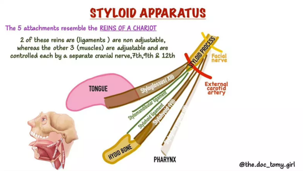
The styloid process is a small, bony, needle-like structure that protrudes from the skull base, and functions as an anchor point for various structures that enable, as well as influence movement of the tongue, pharynx, larynx, hyoid bone, and mandible. These structures are the stylohyoid, stylopharyngeus, and styloglossus muscles as well as the stylohyoid ligament, and the stylomandibular ligament. The styloid process can present as short, long, straight, bent, thin, thickened, etc. That said, most of them are relatively thin, straight, and no longer than 30mm.
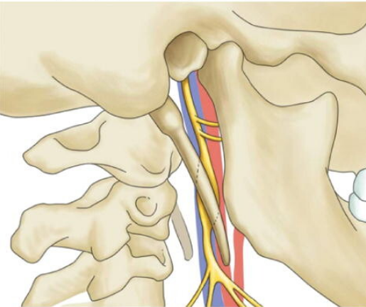
In vascular Eagle’s, although the length of the bone is not of great importance, its angle certainly is, because it will determine the space between the transverse process of the atlas and itself. This is important, because an important neurovascular bundle passes between these two bones, and can become impinged between them. This, ultimately, is what “Vascular” or “Venous” Eagle’s syndrome refers to, and it has nothing to do with the length of the styloid process nor the potential calcification of the stylohyoidal ligament. Due to the latter, in my opinion, it is a completely different pathology from the conventional sense and meaning of Eagle’s Syndrome, and I think its usage causes confusion both amongst patients and clinicians. This is why I have named this condition the “Jugular Outlet Syndrome” (JOS) (Larsen et al., UNPUBLISHED), seeing as the neurovascular bundle that is prone to compression at this site is emerging from the jugular foramen at the bottom of the skull, and also, importantly, because this disorder often involves nerves as well as the internal jugular vein.
Genuine Eagle’s Syndrome
Let us first look at the signs and symptoms of genuine Eagle’s syndrome.
Eagle’s syndrome, in essence, is a condition where soft tissues in the throat and upper anterior neck are exposed to mechanical abrasion or stabbing by an aberrant styloid process or calcified stylohyoid ligament. The most common presentation is with symptoms that resemble glossopharyngeal neuralgia (CN IX), with pain and discomfort in the back of the throat and under the jaw / around the styloid process. But, ES can also irritate the vagus nerve, submandibular parts of the trigeminal nerve, as well as the extra-temporal parts of the facial nerve (Dua 2026, Al-Hashim 2017). In very rare cases, Eagle’s Syndrome can also result in compression, injury, and even dissection of the carotid artery (Maamouri 2022), as well as irritate the autonomic nerves around it.
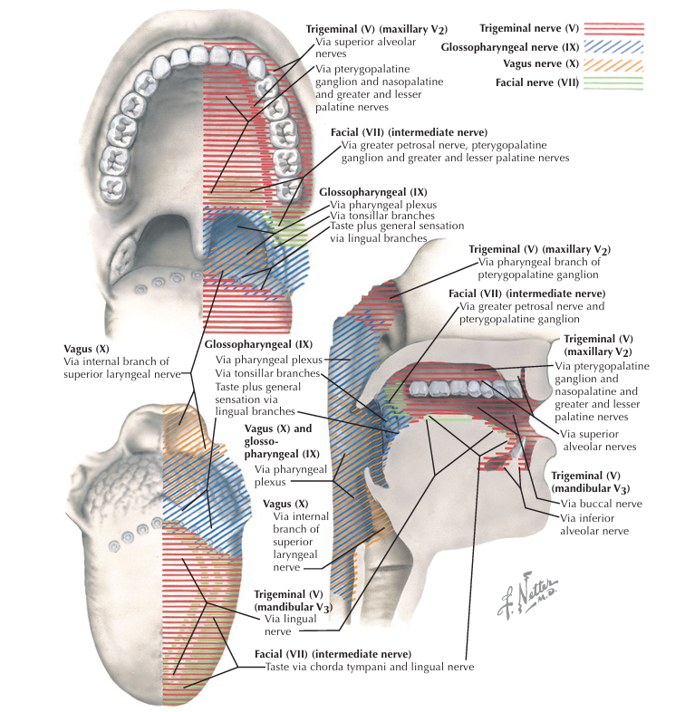
Because of the vast reach of the potential nerves implicated in Eagle’s syndrome, the symptoms can greatly vary. Pain stemming from the glossopharyngeal nerve will usually lead to throat (back of the mouth) pain, but can also cause a feeling of “lump” in the throat, dysphagia, as well as anterior neck pain. Because the glossopharyngeal and vagus nerves are interconnected, compression of one can also affect the other. However, generally, compression of the vagus nerve can lead to throat pain, ear pain, voice hoarseness, dysphagia, chronic dry cough, gastroparesis, lump in the throat sensations (globus hystericus) and diffuse inner / middle ear symptoms. It can also cause eustachian tube dysfunction or patulous eustachian tube. When the facial nerve is affected, pain can arise around the jaw, ear and face. Trigeminal nerve can also cause a myriad of strange facial and head symptoms, but may also include the palate, teeth and tongue, as seen in the illustration below. The tongue can also be affected by the vagus and glossopharyngeal nerves, causing pain, paresthesiae or burning tongue syndrome (Larsen, UNPUBLISHED). Finally, positional compression of the internal or external carotid arteries or their external sheath of autonomic nerves can lead to a myriad of symptoms including transient ischemic attacks, vertigo, and syncope. Pain can also be generated from vascular compression, and is often referred to the supraorbital region (Dua 2026).
The Styloid process is normally no longer than 30mm, measured from the base of the temporal bone. The exact path and location of the various anatomical structures in the throat vary from person to person, and are sometimes a bit smaller, larger, a bit to the left or the right, above or below where the exact same structure is situated in another person. For a normal, asymptomatic person, the styloidal length is short enough to avoid abrasion of adjacent tissues. However, when the tip is longer, bent, or ridden by abrasive osteophytes, then this can be the determining factor for what leads to actual symptom development.
It is important to understand that the styloid process, because it attaches to the temporal bone, moves in synchrony with the skull. The skull and upper cervical vertebra (the “atlas”) offer great mobility with regards to flexion, extension and rotation. The longer the styloid process is, the more likely it is to collide into, and potentially irritate or injure structures during movement. Herein implied is also the notion that movement of the head, and especially certain positions, will be the main trigger for genuine Eagle’s syndrome. It is common to trigger the symptoms with head rotation, flexion and extension, as well as yawning and chewing. When the jaw is involved, this can be confusing, providing potential evidence for a temporomandibular problem or Costen’s syndrome, rather than ES or parallel to ES. Despite being confusing, at-a-glance, the jaw can also be implicated in genuine Eagle’s syndrome because mandibular lowering (mouth opening) may push the vulnerable soft tissues backwards and into the path of the styloid process.
Diagnosis
The diagnostic process is difficult in Eagle’s Syndrome, because the symptoms that arise from this condition can overlap with symptoms from other conditions. For example, patients with Thoracic Outlet Syndrome can frequently present with anterior and lateral neck symptoms that worsen with neck movements, especially rotation, and even the sensation of globus hystericus. However, TOS tends to trigger with arm-loading more so than neck movements, and will generally not see the same level triggering by neck movements as in genuine ES. Most patients with TOS will be reactive to neck movements only in positions that lead to passive tautness or repetitive loading of the scalene muscles, whereas this will be irrelevant for an ES case. Finally, ES cases are not reactive to costoclavicular loading, in stark contrast to the TOS patient. Cervical plexus entrapment syndrome, which I have written sparsely about in other articles on this site, can affect the C1-4 nerves and lead to similar symptoms of throat, submandibular, ear, and facial pains, excluding the cranial nerves. For this condition, isolated and repeated levator scapula stimulation should reproduce the symptoms, something that would not happen in ES. It is therefore crucial to exclude other, potentially compatible pathologies as well as to carefully compare the patient’s symptoms and triggers to what would be expected in a genuine case of ES:
- Involvement of one or more of the cranial nerves (CN 5, 7, 9, 10, 11), but, especially the glossopharyngeal nerve, being the most commonly afflicted
- Provocation of symptoms in certain head/neck positions, or repetitive movements in certain directions of the head and neck
- Abnormal soreness, preferably reproduction of the main symptom upon palpation of the styloid process and tonsillar fossa
- Exclusion of other conditions (this is a difficult topic that exceeds the scope of this article), including JOS! Beware that JOS is a common malady, and that genuine Eagle’s syndrome is rare.
- And, of course, abnormal styloidal length and/or calcification of the stylohyoidal ligament, preferably identified through computed tomography (CT). A CT will also be helpful in determining the space between the styloid and C1 transverse process. A positional CT angiography, where the patient is placed in the provocative position, would also be able to indicate any mechanical insult to the traversing carotid arteries
- An expert radiologist might be able to identify signal changes in one of the cranial nerves through contrasted MRI, but this would require acute inflammation of the respective nerve at the time of imaging (Al-Hashim 2017). Generally, do not expect an MRI to prove the neuropathy; a normal MRI does not disprove ES.
- In rare cases, calcification or injury to the stylomandibular ligament (Ernest’s syndrome)
- If you are predominantly able to elicit the patient’s symptoms during retraction of the head or chin tucking, then JOS would be the prime suspect in contrast to ES
- Genuine ES tends to be one-sided, but it can, in rare cases, be bilateral
Thus, in genuine ES, symptoms should be triggered by palpating around the styloid process’ tip, either orally by the tonsillar fossa or externally just behind the ramus of the mandible. Imaging should show lengthening of the styloid, or calcification of the stylohyoid ligament. Finally, the patient should remark obvious provocation pertaining to head rotations and repeated flexion and/or extension of the neck. Many patients also elicit symptoms by yawning, vigorous speech and tongue extension. Because this can overlap with triggers frequently seen in TMD and Costen’s syndrome, it is important to distinguish the two: Costen’s syndrome and TMD should not be reactive to head movements. If head/neck movements are a repeatable and important trigger for the patient’s symptoms, then a temporomandibular origin of symptoms is less likely. However, if the patient’s symptoms are clearly provoked by jaw use, but do not seem to be particularly influenced by head and neck positioning or movements, then a case of ES is less likely.
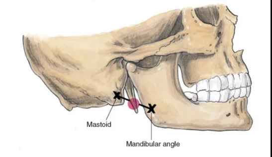
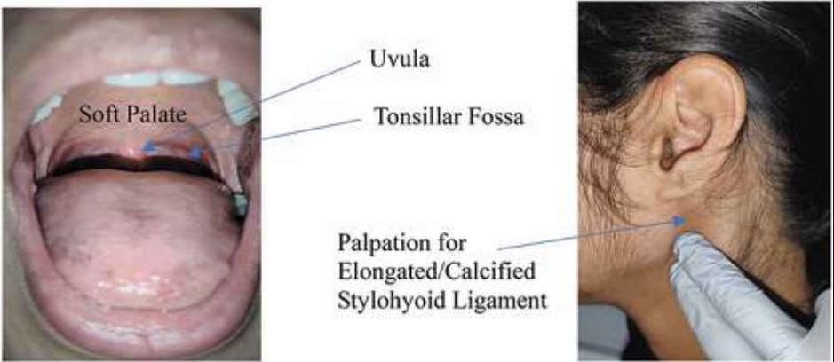
Diagnostic injections for Eagle’s syndrome are often a hit-or-miss and can lead to false negatives in some cases. A failed diagnostic block does not always conclusively disprove of Eagle’s syndrome. Furthermore, a positive response to palpation of the peri-styloidal space or tonsillar fossa will not conclusively distinguish between Jugular Outlet Syndrome and Eagle’s syndrome. However, if a tonsillar fossa injection has an obviously beneficial effect on the patient’s symptoms, especially in the presence of a normal atlanto-styloid interval and a long styloid process or calcified stylohyoid ligament, and the patient’s symptoms trigger with head [and jaw] movements, then you are probably dealing with a genuine case of Eagle’s syndrome.
The treatment for this condition is styloidectomy and resection of the stylohyoid ligament. When diagnosed accurately, successful surgical resection of these structures would be expected to be curative, presuming that no permanent nerve damage has been sustained already, which is rarely the case. Shaving or removal of the C1 transverse process is not necessary nor helpful in genuine Eagle’s syndrome.
Jugular Outlet Syndrome / “Venous” Eagle’s Syndrome
As opposed to genuine Eagle’s syndrome, which is rare, Jugular Outlet Syndrome (aka. Venous Eagle’s Syndrome, Jugular vein compression syndrome, extrinsic jugular compression syndrome, etc) is a common disorder, though many suffer without ever getting diagnosed. The most common set of symptoms associated with this disorder stems from the jugular vein, which when compressed can lead to various symptoms of venogenic intracranial hypertension, such as headache, vertigo, tinnitus, pulsatile tinnitus, migraine, visual derangement, and more. Symptoms can be bilateral or unilateral; both are common. The nerve effects related to JOS are similar to those in genuine Eagle’s Syndrome, although the nerves would be compressed in a different place. This condition has received increasing levels of attention in the medical journals for the last 10 years, but was virtually unknown prior to that. Still, unfortunately, the many recent papers on this topic are generally only known by inner-circle specialists.
JOS / VES is caused by approximation between the styloid process of the temporal bone, and the transverse process of the atlas. This is mainly a postural problem (Larsen 2017, Larsen 2018), but is often also predisposed by a posteriorly angulated styloid process and/or a wide transverse process (Gweon 2011). Gweon et al. found that an atlanto-styloidal interval of approximately 8mm was normal. It is important, however, to understand that this interval depends on the patient’s posture at the moment of imaging, and typically gets worse during flexion and retraction of the head (Larsen 2018, Yoshimura 2026, Pedersen 2015, Seoane 1999). In essence, JOS is a postural phenomenon. Already in 1999, Seoane & Rhoton described intraoperative cerebellar venous hemorrhages secondary to postural IJV obstruction, showing the dramatic potential effect posture has on internal jugular drainage. Ding (2019) found that up to 70% of patients with cervical spondylosis had at least one-sided internal jugular vein stenosis. I have written extensively about the mechanism of this problem in previous articles, and especially in my first atlantoaxial instability article (link).
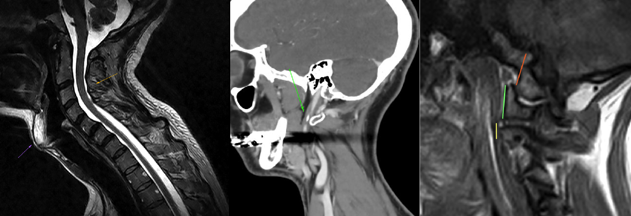
When the head (chin) is drawn down, this draws the anterior head towards the neck and, therein, also draws the styloid towards the C1 transverse process. Likewise, instigating retraction of the head will cause the occipital condyle to translate posteriorly on the C1 in the atlanto-occipital joint, further narrowing the space between these two bony structures. On the other side, “lengthening” of the neck along with slight occipital protraction and extension will generally increase the atlanto-styloidal space. I have videos demonstrating these movements on my YouTube channel (link).
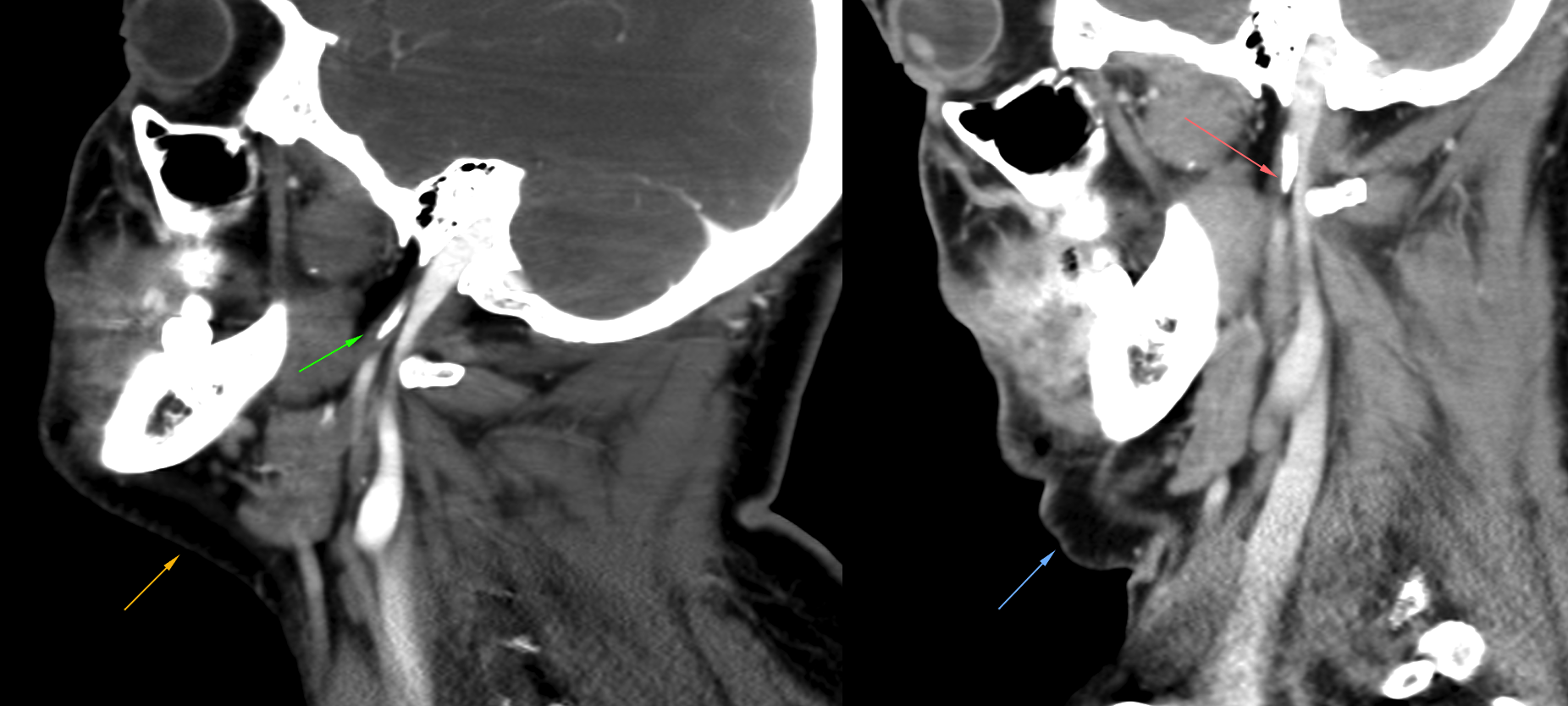
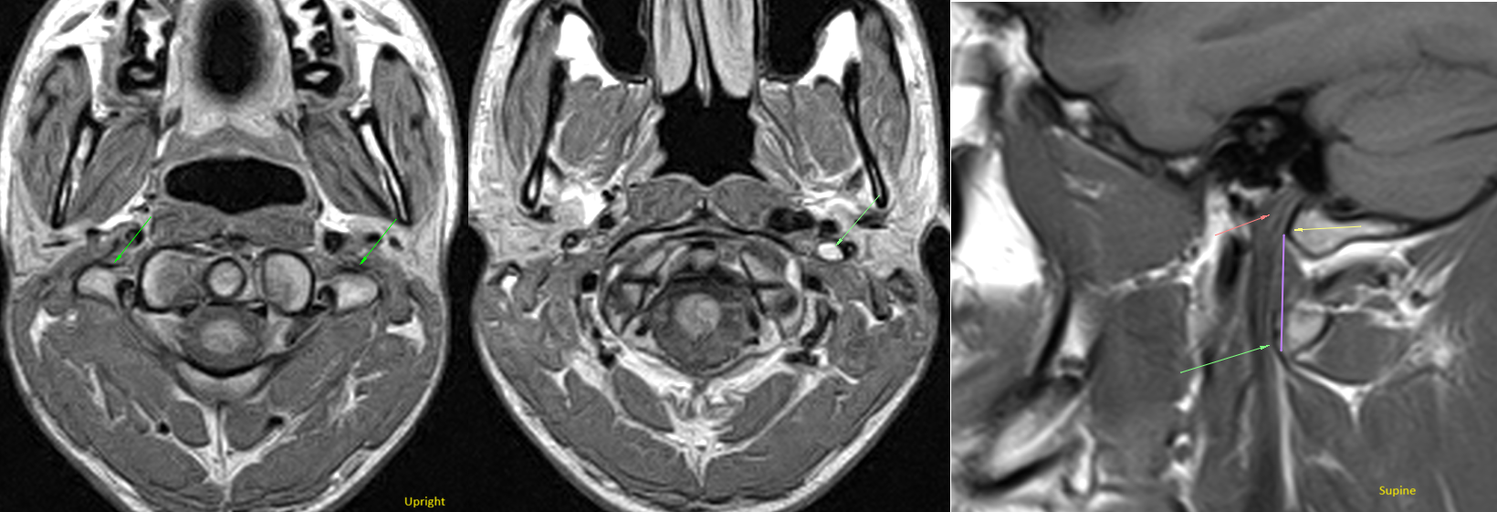
In early and mild cases of JOS, a supine MRI and CT are usually not able to demonstrate any narrowing of the jugular outlet, but it can be seen on an upright MRI of the craniocervical junction. In moderate cases, usually, a supine CT will still be unable to show narrowing, whereas a supine MRI as well as an upright MRI will both demonstrate the abnormality. In severe cases, even the supine CT will show narrowing of the atlanto-styloidal interval. This, again, shows how important postural factors are in the influence of JOS.
The pathomechanism of JOS is as follows:
- A combination of poor posture, over time, and/or unfavorable width of the C1 transverse process or angle of the styloid process leads to approximation between the C1 transverse process and temporal styloid process due to posterior translation of the occipital condyle on the C1 (cf. the illustration above). Some people refer to this condition as craniocervical instability (CCI), but I think that is an overkill statement considering the true implications of CCI, although some of the same mechanisms are involved.
- The bony approximation leads to pinching of the internal jugular vein (IJV) and/or glossopharyngeal, vagus or accessory nerves as they emerge from the jugular foramen and traverse between the two bony landmarks
- The extent of compression is posture-dependent, and usually gets worse in retraction and flexion of the craniocervical junction (ie. the head being pulled back while the chin is tucked), although this does not apply to everyone.
- a) Compression of the internal jugular veins leads to craniovenous outlet deficiency, and thus practically clogs the craniovascular system, leading to craniovascular hypertension (either intermittent or perpetual, depending on severity), increases the risk for cerebral venous sinus thrombosis, and increases the risk for developing elevated cerebrospinal fluid (CSF) pressure and a misdiagnosis of idiopathic intracranial hypertension (IIH). b) Longstanding elevations in CSF and venous pressures can lead to minor spontaneous CSF leaks (Higgins 2015), and cause a very complicated constellation of clinical symptoms, often resulting in the patient’s condition being determined psychogenic or idiopathic.
- Compression of the cranial nerves will lead to similar symptoms as in genuine Eagle’s Syndrome.
- Swelling from overuse or injury, or fatty hypertrophy of the longus capitis muscles can further encroach on the space between the styloid process and the glossopharyngeal and vagal nerves, although it is not readily obvious when conferring with anatomical illustrations. The longus capitis acts as the medial border for the neurovascular bundle, and can result in slight lateral translation of the traversing cranial nerves when swollen. The tiniest change in position can make the difference between being untouched, and being impinged. This especially applies if the styloid process has a medialized angle.
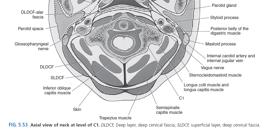
Potential symptoms associated with jugular outlet syndrome:
Craniovascular: Headache, migraine, dysopsia, diplopia, divergence & convergence dysfunction, tinnitus, vestibular dysfunction, seizures, syncope, orthostatic incompetence, impaired cerebrovascular autoregulation, dysarthria, dysphasia, amnesia, ptosis or lid retraction, perceived intracranial pressure, asthenia, optic nerve sheath dilation, papilledema, cerebellar ectopia, cerebral edema (increased volume, no signal change), derealization, depersonalization, depression, Lhermitte’s signs (rare), [secondary] CSF leaks, bradycardia
[Extra-mastoid] Facial nerve: Facial neuralgia, middle ear dysfunction, tinnitus, stapedius myospasm, platysmal dystonia or myoclonus
Glossopharyngeal nerve: Throat pain, globus hystericus (“lump in the throat”), dysphagia, oromandibular dystonia, burning tongue syndrome (also seen in hypoglossal neuralgia)
Vagus nerve: Chronic dry cough, hiccups, ear itches, otalgia, dysphagia, constrictor spasm, eustachian tube dysfunction, patulous eustachian tube, gastroparesis, generalized dystonia or myoclonus, diffuse myoclonic disorders, generalized myoclonic attacks or tremors, complex global pain syndrome (autonomic dysfunction), and cardiac arrhythmias
Accessory nerve: Trapezius or sternocleidomastoid weakness, cervical dystonia or myoclonus (either rotational (one scm or one trapezius affected), protractive (both SCM-affected) or retractive (both trapezius-affected)), accessory n. neuralgia
Diagnosis
Extrinsic internal jugular vein stenosis
Jugular outlet syndrome, as indicated, is a two-headed beast. The venous compression-side of JOS is relatively easy to prove, because it will be visible on MRI, CT and even ultrasound to those who know what they’re looking for. What can be harder to determine is the actual effect of the venous compression, because some patients have excellent, whereas others, poor accessory drainage channels.
The first step is to look for compatible symptoms. Patients with symptomatic IJV stenosis are often struggling with headaches, migraines, fatigue, dizziness and tinnitus. These are the most common symptoms amongst “typical” patients. In the severely afflicted, the aforementioned symptoms in greater severity can be expected, along with other problems as syncope, or neuropsychiatric symptoms such as derealization and depersonalization. These patients are usually worse after being upright for prolonged periods of time, and tend to feel better in the morning. This is because IJV drainage tends to be better when they lie down, and worse when upright — the latter having an accumulative effect.
- Worse in the evening / after a sustained period of being upright
- Reproduction of symptoms (eg. headache or dizziness) after a sustained 15-second Valsalva maneuver
- Reproduction of symptoms after a 1-minute hold of the Queckenstedt’s test
- Worsening after persistent talking (talking, especially vigorous speech, will transiently increase central venous pressures)
- Worsening after bending down to tie your shoes
- Worsening after defecation, especially vigorous defecation
- Worsening with vigorous coughing
On visual inspection, a plain supine cerebral MRI is the best bet. Here you will be able to determine, usually with a combination of axial and sagittal sequences (plain T2-weighted sequences are preferable but not mandatory), whether or not, and to what extent, the internal jugular veins are encroached upon. This alone is not enough to confirm the patient’s cause of symptoms, unfortunately, and follow-up studies are often required. A particularly useful study, though not often used, is a Doppler exam of the internal jugular vein with volume flow measurements. Previous studies have shown that a normal combined (left + right) flow volume in the internal jugular veins is approximately 700-1200mL/min (Özen 2014, Müller 1990, Brunhölzl 1990). Özen et al. also showed that flow rates inferior to 160mL/min were associated with partially occlusive thrombosis, which is interesting. I have personally seen hundreds of patients with flow rates inferior to 160mL/min without thrombosis, but the important take-away here is that sub 160mL/min flow rates are to be interpreted as pre-occlusive flow rates and, as such, highly abnormal.
To get an accurate internal jugular vein flow rate, measurements should be done with appropriate angle correction and gate size at the distal third of the internal jugular vein, at a site where its caliber is as homogeneous as possible. If you measure the flow rates in a segment that is very wide, very narrow, or a site where a lot of caliber undulation is present, then the measurement will not be accurate.
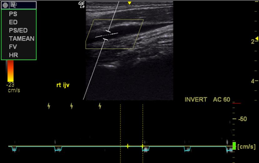
I have seen countless cases where there internal jugular veins appear severely obstructed on imaging, where a doppler exam have shown normal or near-normal flow rates. In such a case, although there may still be restricted flow when upright, it makes it easy to compare the flow rates to the patient’s symptom severity and, coherently, decide whether to proceed with a catheter manometric exam and DSA venography, or move on in pursuit of another differential diagnosis.
Ultimately, a catheter manometric exam is the gold standard for confirmation of diagnosis. What I have found most useful is to look for pressure gradients between the torcula (ie. confluence of sinuses) and the supraclavicular part of the jugular vein, or central venous pressure. Some clinicians get hung up with pre-and post stenotic pressure gradients in the IJV and I have found this to be less useful than expected. According to the literature, a pre-post stenotic pressure gradient of 5mmHg is significant (Fargen 2026). Personally, although I do not disagree with this, I find that looking for gradients between the torcula or SSS and CVP may have improved sensitivity.
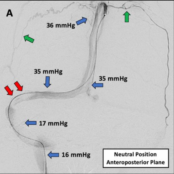
I am also aware that some recent experts and researchers in IJV stenosis have become fond of positional catheter manometry, where the patient is measured in various positions. Personally, although flexion and extension can be useful, I do not find rotational pressure changes very pertinent. The reason for this is that most of these patients feel poorly even in neutral posture, and therefore, if flow is normal in neutral, it is natural to expect something else to be the main driver of their symptoms.
Therefore:
- Look for compatible symptoms (see above)
- Look for compatible triggers (see above)
- Look for IJV narrowing on MRI, or CT (CT will be the last to be positive, suggesting a greater severity)
- Confirm flow rates appropriately through a doppler volume flow exam of the inferior part of the IJV. Total flow should be higher than 700mL/min as a minimum
- Consider a catheter exam in neutral, flexion and extension where CVP, proximal jugular, jugular bulb, left and right tranverse sinuses, torcula, and the SSS pressures are measured. Typically, less than 10mmHg gradients should be seen between CVP and the torcula.
Neurogenic jugular outlet syndrome
For the neurogenic component of Jugular Outlet Syndrome, diagnosis can be harder to conclusively prove. In order to diagnose these patients with reasonable accuracy, it is crucial to understand the potential symptoms, but also pathophysiology and biomechanics of JOS. Even the symptoms in- and by themselves is a difficult topic to manage due to unusually vast array of potential signs. Your first clue is generally going to be throat pain, diffuse dysautonomia (eg. syncope, gastroparesis, or chest palpitations) or dysautonomia / myoclonies that get worse with chin tucking and neck retractions.
For most patients, reiterated, the atlanto-styloidal space will encroach as the head is pulled backwards and/or into flexion (not to be mistaken for NECK FLEXION, as that would be different). Less commonly, the space will narrow upon extension of the neck and head at the same time. Once clinical suspicion is rendered, usually based on symptoms and history alone, then a chin tuck-test can be done.
I use the following approach: Guide the patient into an appropriate (read: terrible) retraction of the head while they gently lead the nose down. If the chin is tucked downward too hard, the neck will start to flex, and the atlanto-styloidal interval will sometimes widen rather than narrow. I then have the patient hold this position for anything from 1-2 minutes depending on how elusive the diagnosis is. In special circumstances, I may have them hold the position longer. Beware that these tests should not be done unsupervised. You are generally looking to elicit the symptoms during the test, but sometimes, as often as 1/3rd of cases, the symptoms may manifest after the test instead. Sometimes several hours later, or even the next day. It is important to be patient and not make premature conclusions and to carefully map out the pattern of symptom manifestation and triggers in each case.
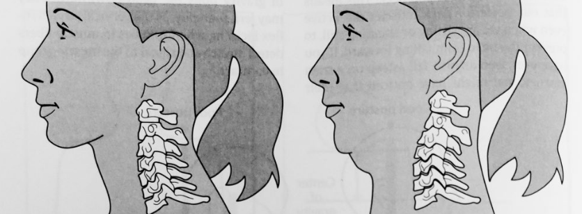
If you can establish that the patient’s symptom is possibly or clearly influenced by narrowing of the atlanto-styloidal interval, then I still recommend to repeat the test a few times. Not on the same day, but over the course of a week. This will ensure diagnostic accuracy and mitigate the chance of misconceptions.
A pitfall to avoid is to get overly hung up on the appearance of the CT. As I have shown, the CT is the last exam to show blatant narrowing of the atlanto-styloidal interval, and you can miss many cases if you fail to trust the clinical data acquired from physical examination. It is not unusual to see IJV stenosis on a supine MRI, but for a later CT to demonstrate normal conditions. This can be confusing, but happens due to the neck wedge that is typically used during a CT examination. The neck/head wedge lifts the patient into slight cervical flexion, spacing the atlanto-styloid interval. An upright MRI, although it does not show the styloid process, will always be the most sensitive study for IJV stenosis. Second up is a plain supine cerebral MRI, preferably with T2-weighted sequence in 1-2mm slices from axial and sagittal perspectives.
Treatment
Surgical
Treatment for JOS is really boxed into surgical and non-surgical cases. Within the surgical group, the addition of venous compression makes things more complicated and often less responsive to mere removal of the styloid process. Let me elaborate.
As you may have grasped by now, reduction of styloidal length (ie. reduction to a normal 20-30mm) or resection of the stylohyoid ligament will do nothing for a case of JOS, because the problem is not abnormal length, but approximation between the styloid and C1 transverse processes. Thus, from a surgical standpoint, the styloid process must be removed well above where the C1 transverse process is situated. I have seen many cases where insufficient removal of the styloid process leaves a stump that just barely clears its previous collision path with the transverse process. The problem here, is that the internal jugular vein will still be kinked between the styloid remnant and the C1 transverse process. Thus, generally, for JOS / VES management, the styloid process should be resected off its base.
Unfortunately many surgeons are reluctant to perform this procedure, because:
- It is an unusual request and most surgeons are not trained in this particular procedure as an Eagle’s syndrome intervention
- They are not familiar with jugular outlet syndrome / VES
- There is a much greater risk for iatrogenic facial nerve damage when attempting to remove the styloid off its base
To my knowledge, Dr. Axon in collaboration with Prof. Dr. Nicholas Higgins (Higgins et al 2017) were the pioneers of these procedures some ten odd years ago. At this point, JOS VES has become a somewhat more known issue, and it is much easier than before to find a surgeon who is willing and competent in removal of the entire styloid process.
Complete removal of the styloid process is expected to cure neurogenic JOS. However, in cases of additional or predominant venous compression and adherent symptoms, even full removal of the styloid process will tend to be inadequate. On post-op imaging, in most of those cases, you will see how the transverse process still acts as an obstacle for the jugular outlet. In milder cases, this will only be visible on upright MRI. But, of course, a mild case has no surgical merit. So, in all surgical cases, the transverse process of the C1 will be visibly anterior and obstruct the jugular outlet horizontally. This problem has caused many surgeons, including the initial pioneers, to perform either anterior shaving or complete removal of the C1 transverse process. Removal of the SP and TP completely will, of course, cure any hint of venous stenosis at the jugular outlet. The problem with this procedure is that it may require tenotomy (cutting the attachment of) several important stabilizing muscle tendons that attach there. Therefore, if a gentle anterior shave can be done without compromising any of the muscle tendons, then this may hold promise. However, removal of the transverse process and tenotomy of the levator scapula, splenius cervicis, rectus lateralis, obliquus inferior & obliquus superior muscles is discouraged in my opinion.
Stenting of the jugular vein is also, generally, not a very viable treatment option, because:
- The stent will be crushed between the styloid and transverse processes (Dashti 2012)
- There is a lot of movement in the segment, predisposing to complications and recurrence, stent dislodgement and potentially grave consequences (Fargen 2024, Liu 2025)
- Even after styloidal removal, the stent will still collide with the transverse process and be prone to failure (Higgins 2017)
- If both the transverse AND styloid processes are removed, there is no longer any need for a stent
Therefore, in most cases, a complete resection of the styloid process can be considered if the patient fails conservative management, or whose symptoms warrant surgical intervention. In my experience, patients who demonstrate very narrow atlanto-styloid intervals on CT are harder to treat conservatively, and may benefit from surgical removal of the styloid process if their symptom severity is consistent with such a choice. Less than 3mm space on a CT is often to be interpreted as “severe”. As mentioned earlier, if you find at least a 4-5mm space on CT, then the likelihood of favorable outcomes through appropriate conservative management is higher.
As shown, styloidectomy alone is often not enough to completely decompress the internal jugular vein. In fact, I have seen masterfully done styloidectomies with additional transversectomy, where pre- and post-op imaging shows complete resolution of the stenosis. However, the very same patient still demonstrated 80% narrowing of both internal jugular veins on an upright MRI, once again showing how important the postural aspect is in the management of this condition. Instead of performing styloidectomy and transversectomy, my recommendation is to leave the transverse process alone, unless you are able to just shave a bit off its anterior aspect without damaging any of the tendons, and to proceed to post-op conservative therapy afterwards. This will be discussed next.
Conservative / Post-operative approach
Appropriate conservative management of this condition is warranted with- or without surgery. As we have seen, surgery alone will generally not lead to complete liberation of the jugular outlet. I therefore recommend specific approaches for neck and head posture, along with specific muscle strengthening protocols that aim to stabilize the upper neck, and liberate the jugular outlet.
The first step is the postural correction. This change also effects atlanto-styloidal spacing; decompression of the jugular outlet. “Lengthening” of the neck, along with gentle protraction and extension of the head will space the atlanto-styloidal interval.
Mechanically, in isolation, it is that simple. Unfortunately, two common, and significant setbacks often present themselves when commencing treatment for this condition.
Firstly, appropriate decompression requires excellent function of the deep neck flexors (especially the longus capitis), in addition to the levator scapula and suboccipital muscles. The latter two are less reactive to postural changes, but, alas, the deep neck flexors can be extremely reactive to overuse. After neck injury (eg. whiplash), or years of poor posture, especially when combined with stress and “clenching”, can cause severe detriment to the longus capitis. The longus capitis is a noteworthy structure due to its crucial impact on craniocervical stability and alignment, and its adjacent sympathetic chain. Overloading tends to occur very easily, and this can lead to many problems. Firstly, longus capitis and longus colli overloading leads to amplified (but intermittent) weakness, and this causes a sensation of “wobble-head” (a symptom many mistake for CCI). Further, the overloading leads to muscle swelling, which further decreases the nerve space in the triangle between the longus capitis, styloid process, and C1 transverse process. Finally, overloading and subsequent inflammation of the longus capitis can lead to entrapment of its adjacent traversing sympathetic chain, and therein a myriad of potential problems:
- Tinnitus
- Headache
- Migraine
- Dysopsia
- Syncope
- Asthenia
- Orthostatic incompetence and POTS
- Deranged cerebrovascular autoregulation
- Eustachian tube dysfunction
- Patulous eustachian tube
Over the years, I have had several patients overdo this and get quite ill. It is imperative to listen to your body and give it time to accommodate the changes. It is not unusual, for a patient who has been “hinging” on their necks for decades, to spend a couple of years restoring a proper neck posture, including proper rehabilitation of the deep neck flexors.
Furthermore: In many of these cases, even if overloading of the longus capitis is successfully avoided, the patient may find themselves in a state where the nerve is “comfortably numb”, with the lack of a better term. In these cases, you will find that increasing the compression of the nerve causes a delayed rather than instantaneous worsening of their symptoms. This is crucial to understand, as failure to do so may easily lead to misinterpretation and misdiagnosis. In these cases, despite having confirmed worsening (over time) through repeated provocative chin-tuck tests, many will find that decompression also leads to increased pain generation. This phenomenon occurs because the nerve has been compressed for so long, often years, and is no longer working properly. When decompressed, its function slowly returns, and during this initial stage, severe symptom provocation can occur despite the patient doing everything correctly, while the nerve heals. Of course, you always want to double- yes, triple check that you are indeed doing everything correctly, not merely basing your conclusions on assumptions. Either way: I call this process “unnumbing” of the nerve. This very frequently leads to patient-discouragement and getting cold feet about the protocol, fearing misdiagnosis or that are on the wrong path.
It is important to understand these pitfalls prior to starting treatment so that you can best prepare yourself for what’s ahead. Patients with lower level symptoms are less prone to these complications, but patients with severe and longstanding symptoms are much more likely to go through the exact problems that I have elaborated above.
Exercises
Also essential, but less prone to complications, are the exercises for the muscles that encourage anterior gliding of the skull on the upper neck. This, along with the postural correction is what encourages and maintains spacing of the atlanto-styloidal interval. The suboccipital and levator scapula muscles are the most important components in the exercise regimen. I strongly discourage adding additional longus capitis and longus colli exercises, because it will already be very taxed from the postural work. The postural work alone is more than enough for the deep neck flexors, and it is in your best interest to take my word for that.
I recommend strengthening the levator scapulae and suboccipital muscles for one set until moderate fatigue, twice per week.
I recommend that you read my first atlas article (linked earlier) for more elaborated explanations of upper neck rehabilitation.
Final thoughts
I have a large paper on this topic that I have been wanting to finish since 2019, but since I still haven’t gotten that done, then this will be the second best, for now. Jugular Outlet Syndrome is a common disorder, compared to the rare convenetional Eagle’s syndrome. And, as you surely have inferred, the two conditions have different pathophysiological mechanisms.
For conventional Eagle’s Syndrome, shortening of the styloid process along with potential resection of the stylohyoid ligament (if calcified) will have curative effects, presuming that no permanent nerve injury has been sustained. This is generally not the case, and many ENT departments around the world carry out these procedures, although it can be difficult to be diagnosed. It is rare, after all.
Jugular Outlet Syndrome, aka. “Venous Eagle’s Syndrome”, on its side, is poorly known, and there are few medical centers trained in its management, despite it being a very common problem. The vast majority of patients afflicted are never diagnosed, or find it extremely hard to get diagnosed. For many years, and still in most places, internal jugular venous compression at the C1 level is considered a developmental anomaly rather than pathology. It is crucial to understand that this condition mainly develops due to poor posture and weak upper cervical musculature, over time. But it can also be instigated after a head/neck injury, eg. after a whiplash incident.
Genuine Eagle’s syndrome is a surgical problem, but Jugular Outlet Syndrome can be managed successfully through appropriate physical therapy protocols if the patient is patient and diligent. Compliance is absolutely mandatory if you are to see any results, especially in advanced cases. But, again, for an advanced case, then combination therapy of styloidectomy followed by these physical therapy protocols may be the best option. And yes, it is possible to have both at once, which is why taking the styloid process off its base is generally a good idea in the first place if surgical intervention has already been decided.
References
- Karapepera VR, Skarmoutsou AAM, Tziouris D. Eagle’s Syndrome: A Fortunate Discovery in a Symptomatic Patient. Cureus. 2024 May 27;16(5):e61181. doi: 10.7759/cureus.61181. PMID: 38933649; PMCID: PMC11200315.
- Dua A, Mohseni M. Eagle Syndrome. [Updated 2026 Mar 25]. In: StatPearls [Internet]. Treasure Island (FL): StatPearls Publishing; 2026 Jan-. Available from: https://www.ncbi.nlm.nih.gov/sites/books/NBK430789/
- Larsen et al. Jugular Outlet Syndrome (JOS): A common sequela of chronic craniocervical dysfunction. UNPUBLISHED ARTICLE
- Shankland WE 2nd. Ernest syndrome as a consequence of stylomandibular ligament injury: a report of 68 patients. J Prosthet Dent. 1987 Apr;57(4):501-6. doi: 10.1016/0022-3913(87)90025-4. PMID: 3471963.
- Al-Hashim M, Al-Jazan N, Abdulqader A, Al-Ghamdi M. Eagle’s syndrome with facial palsy. J Family Community Med. 2017 May-Aug;24(2):128-130. doi: 10.4103/jfcm.JFCM_134_16. PMID: 28566979; PMCID: PMC5426105.
- Maamouri R, Ouederni M, Oueslati Y, Mbarek C, Chammakhi C, Cheour M. Acute Painful Horner’s Syndrome Revealing Eagle’s Syndrome: A Report of Two Cases. Neuroophthalmology. 2022;46(4):244-247
- Higgins N, Trivedi R, Greenwood R, Pickard J. Brain Slump Caused by Jugular Venous Stenoses Treated by Stenting: A Hypothesis to Link Spontaneous Intracranial Hypotension with Idiopathic Intracranial Hypertension. J Neurol Surg Rep. 2015 Jul;76(1):e188-93. doi: 10.1055/s-0035-1555015. Epub 2015 Jun 12. PMID: 26251803; PMCID: PMC4520985.
- Udhawani, N. S., & Hoover, D. L. (2024). Differential screen and treatment of sternocleidomastoid syndrome versus eagle syndrome: a case report. Physiotherapy Theory and Practice, 40(5), 1072–1082. https://doi.org/10.1080/09593985.2022.2144560
- Larsen, K. Atlas joint instability: Causes, consequences and solutions. Web article, 2017. Available from: https://mskneurology.com/atlas-joint-instability-causes-consequences-solutions/
- Larsen K. Occult intracranial hypertension as a sequela of biomechanical internal jugular vein stenosis: A case report. Anaesth Pain & Intensive Care 2018;22(2):238-242
- Gweon HM, Chung T, Suh SH. Evaluation of the Cause of Internal Jugular Vein Obstruction on Head and Neck Contrast Enhanced 3D MR Angiography Using Contrast Enhanced Computed Tomography. JKSMRM 15:41-47(2011)
- Yoshimura M, Narui M, Kobayashi K, Koh S, Iwasaki S, Naitou K. Styloid-C1 internal jugular vein compression associated with spontaneous cervical epidural hematoma: A case report. Surg Neurol Int. 2026 May 22;17:296. doi: 10.25259/SNI_159_2026. PMID: 42232413; PMCID: PMC13224268.
- Pedersen SSH, Andresen M, Jørgensen AL, Christoffersen DH, Juhler M. Relationship between flexion of the neck and changes in intracranial pressure. Fluids Barriers CNS. 2015 Sep 18;12(Suppl 1):P39. doi: 10.1186/2045-8118-12-S1-P39. PMCID: PMC4582200.
- Seoane E, Rhoton AL Jr. Compression of the internal jugular vein by the transverse process of the atlas as the cause of cerebellar hemorrhage after supratentorial craniotomy. Surg Neurol. 1999 May;51(5):500-5.
- Ding JY, Zhou D, Pan LQ, Ya JY, Liu C, Yan F, et al. Cervical spondylotic internal jugular venous compression syndrome. CNS Neurosci Ther. 2019;00:1–8.
- Özen Ö, Ünal Ö, Avcu S1. Flow volumes of internal jugular veins are significantly reduced in patients with cerebral venous sinus thrombosis. Curr Neurovasc Res. 2014 Feb;11(1):75-82.
- Müller HR, Hinn G, Buser MW. Internal jugular venous flow measurement by means of a duplex scanner. J Ultrasound Med. 1990 May;9(5):261-5.
- Brunhölzl C, Müller HR. [Doppler sonography measurement of jugular vein blood flow]. Vasa. 1990;19(1):26-9.
- Fargen KM, Midtlien JP, Margraf CR, Hui FK. Idiopathic intracranial hypertension pathogenesis: The jugular hypothesis. Interv Neuroradiol. 2026 Oct;32(5):2198-2207. doi: 10.1177/15910199241270660. Epub 2024 Aug 8. PMID: 39113487; PMCID: PMC11571588.
- Fargen KM, Midtlien JP, Margraf CR, et al. Cerebral venous pressures, sinus trans-stenosis gradients, and intracranial pressures are dramatically augmented by head positionJournal of NeuroInterventional Surgery 2025;17:1120-1126.
- Higgins JN, Garnett MR, Pickard JD, Axon PR. An Evaluation of Styloidectomy as an Adjunct or Alternative to Jugular Stenting in Idiopathic Intracranial Hypertension and Disturbances of Cranial Venous Outflow. J Neurol Surg B Skull Base. 2017 Apr;78(2):158-163. doi: 10.1055/s-0036-1594238. Epub 2016 Nov 23. PMID: 28321380; PMCID: PMC5357221.
- Dashti SR, Nakaji P, Hu YC, Frei DF, Abla AA, Yao T, et al. Styloidogenic jugular venous compression syndrome: diagnosis and treatment: case report. Neurosurgery. 2012 Mar;70(3):E795-9. doi: 10.1227/NEU.0b013e3182333859
- Fargen KM, Midtlien JP, Belanger K, Hepworth EJ, Hui FK. The Promise, Mystery, and Perils of Stenting for Symptomatic Internal Jugular Vein Stenosis: A Case Series. Neurosurgery. 2024 Aug 1;95(2):400-407. doi: 10.1227/neu.0000000000002891. Epub 2024 Mar 13. PMID: 38477595.
- Liu X, Wang P, Yang K, Zhu S. Complication of stent displacement from the internal jugular vein to the pulmonary artery after jugular vein stenting in the treatment of intracranial hypertension: a case report. BMC Neurol. 2025 Nov 18;25(1):473. doi: 10.1186/s12883-025-04484-y. PMID: 41254519; PMCID: PMC12625422.
